#epicardium
Explore tagged Tumblr posts
Visit Tumblr Blog
Explore Tumblr blogs with no restrictions, modern design and the best experience.
Last Seen Tumblr Blogs


Fun Fact
The Tumblr office adopted Tommy, an 11-year-old Pomeranian.
Text
Cardiac
Epicardium
The outermost layer of the heart, mesothelial cells and fat, a cushion of protection with TOM written on it in sharpie. He joined my class in Grade 6, chisel-tipped and quiet and an easy choice when asked which Boy I Liked. Which Boy I had a Crush on. That was the year my mom drew eyelashes on me with thick black pencil before a play where I was meant to be beautiful. I wasn’t, but I tried to make myself fit into a beautiful shape, just as I tried to fit the indent in your couch cushions, the foot of your bed, the pillows on the floor, a burrowing owl who would say the right names, make the right choices, pretend I knew who was Hot and who was Not, pretend I knew which house was Tom’s, that I walked past it on my way home from school with my heart like an electric peach, so bright he could see it from his bedroom window.
Brian was the Backstreet Boy I chose from thin air, bad answer, should’ve picked Nick, but at least I didn’t say Kevin, sorry Kevin, he seems like a pretty great dad these days, maybe we all chose wrong.
You would peel apples and read the future in the shape they left on the floor: what is the first letter of your husband’s first name? Light candles and ask the ouija board ‘does he like me? Does he love me? Is he the one?’ Commune with the spirits just to beg them for love stories, and spin a globe and close your eyes and put your fingers down to find out where we’d meet the man-of-our-dreams.
But I wasn’t dreaming about anyone.
My dreams were glass and silver, like the colour of the only eyeshadow my mother owned, brushed powdery and stale up to my eyebrows in a play where I had to sing and a prince had to fall ruinously in love with me in front of everyone I knew. I shone pale blue with shame, and that night, I was the only one at your house who didn’t know the macarena - sat in your basement, watched you dancing, like I was looking into an aquarium filled with strange fish. Later, you’d teach me the steps, like you taught me to fold paper into those fortune-telling finger games, salt-cellar, snapdragon, pick a colour, pick a number, 1-2-3, who will I marry, who will I marry, who will I marry. You put on “Kissing You” by Des’ree, told us to listen in silence and think about the boys we loved, and I wore my longing like a mask that didn’t know it was a mask. Thank you Tom, thank you Brian and Robert and Adam, thank you every boy who let me hold his name in my mouth like an ice cube. The letters burnt my tongue, but at least my mouth wasn’t empty.
Myocardium
The thickest of all three layers, muscle that makes the heart contract, lets it beat beat beat like a kickdrum. I told my first girlfriend that I’d been in love with my best friend growing up, but it wasn’t true, it was just a rhythm I wanted to replicate, to awkwardly dance to. I’d seen the movies and I thought all gay kids had to say it, like it was a shared purple ache in the flesh of us, a thumbprint on a plum. I wanted to feel that bruising early love like everyone told me I should, but it wasn’t like that with us. I wasn’t lying awake looking at the hair on your face, the fascinating black sideburns that you shaved off, like you shaved off the hair on your arms, like I did too. It wasn’t like that, and the night we said we would travel the world together after college, wouldn’t get married or have children, was also the night you said you were glad you didn’t have any gay friends, and remember that book we found at the second-hand store? The air was drowned with dust and Loving Someone Gay shocked us out of papery silence, made you laugh so much that I laughed too, and then I took that book and rolled it up and shoved it down my throat, got paper cuts under my skin, shredded my trachea like tissue paper wrapped around a present.
I wasn’t carrying any torches. Not even a candle or a match.
Your fingers were never in my hair as you pinned it back, you never leaned in and pressed an eyelash to my cheek so that I could wish on it. I wouldn’t have let you touch me. My face my hands my hair, I hated being touched, cringed away from it like a shameplant, and the not-wanting felt almost worse than not-being-wanted. Felt lonely, always the first person awake in your silent basement, bodies scattered like petals on the floor all around me. There was nothing I could do but wait, wait and read your parents’ headache-coloured paperbacks, Louis L’Amour and Danielle Steele, Christ, I hated them, but I would still sit there, paging through Haunted fucking Mesa or whatever, counting down the minutes on the clock, waiting waiting. Sometimes I would hear your father praying in the kitchen but it never woke you up, and I wanted to ache like Courtney Love ached, wanted to feel anything at all except bored and choking on paper, I wanted a drumbeat underneath my skin but it was all silence and darkness and purple muscle, and your father kept praying, ringing bells like they were birds, and I kept waiting to hear music.
Endocardium
Before the world ended, you pressed play on a discman and flooded my life with the Cranberries.
Before the world ended, I looked up at the ceiling and found it strung with hanging lights, each one of them a city in between us. Before the world ended, I asked you if people could yearn in their thirties, if that was allowed, and I didn’t know the steps to this dance but maybe you could teach them to me. Suddenly there was an electric peach in my mouth and it was shining through the spaces between my clenched teeth, anyone could see it, even you. I thought my skin was too thin, my bones too brittle, thought anything I felt would tear through me or grind me down, and maybe it still will but I’d let you press our hips together, iliac crest to iliac crest, let you paint Valentines in red against my lips and mermaids on my cheekbones, let you braid my unbraided hair. Even if it meant you had to touch me
I would let you make me over.
Sometimes the distance feels less like miles, more like inches. Like it could be the space separating your eyelashes from the tip of my nose, could be a hand’s width on a sleeping bag where I’m still awake because you’re lying next to me (and we watched an awful movie with Drew Barrymore in it.) Before the world ended, everything was cold metal and antiseptic, we were hunkering down for the longest winter, planning, preparing, frantic, scared, surviving, sick and I was still the first person awake in a strange basement but suddenly finally now of all the goddamn times I was waking up watermelon-flavoured. My mouth was hard candy, glossy-sweet, and suddenly finally now I was the girl with the most cake, I was peony season, I was Nick Drake and the whole moon shining and the whole sun rising I was pink pink Pink PInk PINK.
#prose poem what I wrote#slumber party zine#it's been the longest of years#this was so much fun to be a part of and I got to stare into the heart-shaped void for a bit#thanks to the mods and editors and contributors for being the loveliest#zines on tumblr
36 notes
·
View notes
Note
You asked for short blurbs! Here are your blurbs!
-Tav hates the wine that Raphael picked for their dinner/date
-The Crown of Karsus doesn't fit between Raphael's horns
-Raphael receives a human heart as a valentine from Tav
Thank you for the prompts @punderdome! I absolutely adore the heart one, and since I'm in no way equipped to write anything even slightly funny (which I think the other two prompts definitely require), I've gone with that one. Hope you enjoy it <3
A gift
The heart is rather mundane. Dull. A fixture both in hell and his house.
Still. It is pretty, all flushed and strong—still beating in his clawed hand, no doubt through magic.
A gift.
That is what Korilla called it. From the hero of Baldur's Gate. His little mouse.
Raphael brings it to his mouth and does what he does best. He consumes. Devours. Teeth dig into the epicardium, through the thick myocardium until they pierce the endocardium, all as blood spills out and over his lips, down his chin—scarlet and bright and fresh.
He moans at the taste. His teeth and tongue works at the organ until every bit has been chewed and swallowed. He can picture it, a warm corpse staining Tav's hands just as he has stained her soul. It only makes him savour it more.
A single, unbidden thought worms its way into his mind—obnoxious and intrusive. Unbecoming.
If only it was hers.
One day, he tells himself. He will not be denied.
#raphael bg3#raphael baldur's gate 3#raphael x tav#raphael x reader#bg3#baldur's gate 3#raphael the cambion#writing
40 notes
·
View notes
Text

Blessed is the cardiac striated muscle tissue that contracted out of Love for us. Blessed is the left ventricle which allowed the blood of the Saviour to reach every part of His body, and blessed is the aorta that branched out to take it there. Blessed is the Vena Cava that gathered all that most holy blood to bring it back to the Heart, and blessed are the right atrium and ventricle for pumping it into the lungs. Blessed are the pulmonary arteries that took it to receive the oxygen it needed for this wondrous, mercyful, painful, incomprehensible task of reconciliation. Blessed are the pulmonary veins, which brought this revitalized blood back to the left atrium, so that it could once more run through His body and give Him the strenght He needed purely because He chose to need it. Blessed are the valves that opened and closed when necessary to allow this process, and blessed are the tendinous cords and the sinoatrial node that made it rhythmic. Blessed are the coronary arteries, which supplied His very Heart with the nutrients demanded to keep It alive and beating. Blessed is the pericardium, which filled with water and blood at the Sorrowful Passion and blessed is every layer - epicardium, myocardium and endocardium - that was ripped open by a spear. Blessed is the Heart of Our Lord, which burned with love for the littlest of creatures. And blessed is every single cell, electric impulse, and extracellular matrix that permitted It to se Its mission through.
(pride flags version)
#my post#my art#sacred heart#sacred heart of Jesus#sorry for any innacuracies! I drew it based on my anatomy textbook but hm. I’m not that good egsjgsjs#sorry guys drawing coronary VEINS was where I drew the line
41 notes
·
View notes
Text

Heart-wrapping Lymphatics
The heart's system of lymphatic vessels transports immune cells and is involved in fluid regulation. This study reveals that these vessels develop in the epicardium – one of the heart's membranous coverings, and that they're initiated by signalling of proteins called VEGFC and D
Read the published research article here
Image from work by Ester de la Cruz and colleagues
Cardiovascular Regeneration ProgramCentro Nacional de Investigaciones Cardiovasculares (CNIC) Madrid, Spain
Image originally published with a Creative Commons Attribution 4.0 International (CC BY 4.0)
Published in EMBO Reports, March 2025
You can also follow BPoD on Instagram, Twitter, Facebook and Bluesky
4 notes
·
View notes
Text
thursday snippet (even though it's not thursday anymore)!!
thank u @sugarsnappeases for the tag<33
you're getting a little piece of the upcoming chapter of east of eden, told from pandora's point of view<3
(the 'evan' she's talking to is her reflection btw)
She hasn't had the chance to wash her hands yet, they still feel filthy. To escape this, she swings into one of the bathrooms on her way; letting the water run until it feels warm enough to scold, sticking her trembling fingers into the ruthless stream from the tab and keeping them there until she can't feel them anymore, it's become quite a routine these days. She leans on the sink when she reaches to turn off the tab with her elbow. Her fingers are red from the throbbing heat of the water, and they dig into the edge of the sink like the smooth, pale porcelain counts as a human touch if she wants it to badly enough. She imagines feeling a pulse beating beneath the glazed surface, imagines clinging to it until she could swear she feels it in her fingers. At last, she recognizes the feeling of the tear that rolls down her cheek silently. She lets it run for a moment, allows it to hurt, before its path curves, and it trickles down into the corner of her mouth. She sticks her tongue out to catch it; giggling, more tears start to fall. As she looks up, fingers digging into the cold porcelain of the sink, she finds Evan in the bathroom mirror's reflection, teary-eyed and laughing back at her through pale eyebrows. She sniffs. The lamp from the ceiling drags harsh shadows under her brow bone, shields her eyes from the warm light. "One day, Evan," she states, eyes glued to her reflection, "Tom Riddle is going to die." She swallows. "He's going to die, and it won't be me who killed him." A soft click sounds from her tongue, a subconscious habit that she picked up from Evan years ago. She leans closer to the mirror's blank surface. "It doesn't matter to me. I don't care if he's dead or alive; I'll carve his heart straight out of his chest, and I'll keep it in a plastic bag until I reach California." She imagines Evan looking at her, a long look that drags out for minutes before he blinks. The heart, he will know about; he will know, instinctively, that they have three beating hearts, now. There will still be guts spilled up to Pandora's elbows; she'll be squeezing the epicardium a hundred and twenty times a minute just to keep it beating through her travel, and when she hands it to Evan, he will put it in a glass jar of liquid nitrogen and place it on her nightstand with a proud smile. "Hello, sister," is what he'll say. "Welcome back to California."
No pressure tags to @sophyayayayaya (dunno if you want the thingie you showed me to see the light of day or not but if you'd like) and also @grimsneverendingfuneral because i'm actually obsessed with your rosekiller snippets 🤭🫀
#'it's unedited' i chant to myself in the mirror like anyone's listening😭#LISTEN fallen angel pandora#(the alexandre cabanel painting i mean)#pandora said there will be guts spilled up to her elbows and i felt that#someone sedate this safety hazard of a woman#pandora rosier#east of eden tag
10 notes
·
View notes
Text
Anatomy of The Heart - The First Time Mother Anatomy of The Heart - The First Time Mother The heart is a crucial organ responsible for pumping blood, supplying oxygen and nutrients, and removing waste from the body. It lies within a double-walled sac known as the pericardium, which shields and decreases friction when it contracts and relaxes. In this sac is fluid that cushions the heart, reducing potential damage from impact or movement. The heart wall is composed of 3 layers (Chruscik 2021). The deepest layer, known as the endocardium, gives a smooth lining to inner chambers and valves so that blood can flow through it without much friction. The middle coat, the myocardium, is muscular tissue that is responsible for producing power to pump blood. The outer layer, called the epicardium, contributes additional protection to the heart. Internally, the heart contains four chambers: the atria (right and left) at the top, which receive blood, and the ventricles (right and left) at the bottom, which pump blood out of the heart. The right side of the heart deals with deoxygenated blood, while the left handles oxygenated blood (National Heart, Lung, and Blood Institute, 2022). Valves between chambers ensure one-way blood flow: the tricuspid valve separates the right atrium and ventricle, and the mitral valve separates the left atrium and ventricle. Additionally, the pulmonary and aortic valves control blood flow as it exits the heart from the right and left ventricles. A thick muscular wall called the septum divides the heart into left and right sides, preventing the mixing of oxygen-rich and oxygen-poor blood. One of the most important divisions in this system is the division needed for blood circulation. The apex of the heart, located at its lower point, directs the pumping action and helps maintain circulation efficiency. Major blood vessels attached to the heart include the aorta (carrying oxygenated blood from the left ventricle to the body), pulmonary arteries (delivering deoxygenated blood from the right ventricle to the lungs), pulmonary veins (transporting oxygenated blood from the lungs to the left atrium), and the vena cava (returning deoxygenated blood from the body to the right atrium). In Laura’s baby’s case, the diagnosis was a ventricular septal defect (VSD), a congenital condition where there is an opening in the septum between the ventricles. With VSD, oxygen-rich blood from the left ventricle can mix with oxygen-poor blood in the right ventricle (Dakkak & Oliver, 2023). This reduces the oxygen content reaching the body and makes the heart work harder to pump enough oxygenated blood. Symptoms can include easy fatigue, as Laura observed during feeding and cyanosis (a blue tint around the lips or tongue) due to low blood oxygen levels. VSD may also be identified through a heart murmur, which occurs due to turbulent blood flow in the heart. Regarding Rina's attitude as a registered nurse encountering this situation, her response was highly appropriate and professional for several reasons. First, she recognized the serious nature of the symptoms Laura described. Cyanosis and feeding difficulties in an infant are red flag symptoms that require immediate medical attention, as they can indicate serious cardiovascular problems. Second, while Rina conveyed the situation's urgency, she maintained a supportive and empathetic approach. When she noticed Laura becoming distressed, she quickly adjusted her communication style to provide reassurance while emphasizing the importance of seeking medical care. Rina's intervention proved crucial, leading to a timely diagnosis and treatment plan for the baby. Her professional knowledge and quick recognition of potentially serious symptoms helped expedite the medical evaluation. The cardiologist's subsequent confirmation of a ventricular septal defect validated her initial concerns and demonstrated that her urgent response was warranted. References Chruscik, A., Kauter, K., Windus, L., Whiteside, E., & Dooley, L. (2021). Fundamentals of anatomy and physiology. University of Southern Queensland. Dakkak, W., & Oliver, T. I. (2023, January 16). Ventricular Septal Defect. Nih.gov; StatPearls Publishing. https://www.ncbi.nlm.nih.gov/books/NBK470330/ National Heart, Lung, and Blood Institute. (2022). How the heart works - how blood flows through the heart | NHLBI, NIH. Www.nhlbi.nih.gov. https://www.nhlbi.nih.gov/health/heart/blood-flow Read the full article
0 notes
Text
Anatomy of the Heart: Understanding the Structure and Function of This Vital Organ
Explore the intricate anatomy of the heart, the powerhouse of the circulatory system. This guide delves into the heart's four chambers—right atrium, right ventricle, left atrium, and left ventricle—along with the critical valves that ensure proper blood flow. Learn about the three layers of the heart wall: the epicardium, myocardium, and endocardium. Discover the role of coronary arteries in supplying blood to the heart itself and understand the electrical system that regulates heartbeat. Whether you're a student, a healthcare professional, or simply curious, this overview will enhance your understanding of how this remarkable organ works to sustain life.
0 notes
Text
It goes without saying that the heart is the most essential organ in the human body. However, did you ever think of the protective casing that takes care of the heart? No, right? The pericardium is the sac that works like a protective casing for the human heart. It ensures that the circulatory system works seamlessly by working with organs like the septum that prevent the mixing of oxygenated and deoxygenated blood during circulation and valves that prevent the backflow of blood in the heart in harmony.
The pericardium has a multitude of roles to play in the body. Circulation is only a single example, but no function of the human heart would be possible without the presence of the pericardium. It is an under-talked face of the human heart that needs more discussion among healthcare enthusiasts and clinicians. Therefore, this blog will do the needful for you.
Here, we will discuss all facets of this protective layer of the heart- pericardium. We will talk about how the pericardium plays a vital role in all functions of the heart. Furthermore, we will discuss pericardial conditions and their clinical implications. So, by the end of this blog, you will be educated about this under-discussed aspect of heart health entirely.
What Is The Pericardium?
The pericardium is a double-walled sac that encases the heart and the roots of the great vessels, consisting of two main layers:
Fibrous Pericardium: This tough, outer layer is composed of dense connective tissue. It anchors the heart to surrounding structures like the diaphragm and sternum, preventing excessive movement during bodily activities.
Serous Pericardium: This inner layer is divided into two sub-layers:
Parietal Layer: Lines the inner surface of the fibrous pericardium.
Visceral Layer (Epicardium): Directly covers the heart muscle.
Between these two sub-layers is the pericardial cavity, which contains a small amount of lubricating fluid that reduces friction as the heart beats.
Functions of the Pericardium
There are more functions of the pericardium than you would imagine. It helps in numerous activities of the heart, ensuring that the harmony between the heart and other organs of the human body remains undisturbed. The following is a list of all the functions of the pericardium:
1. Protection and Support
The primary function of the pericardium is to protect the heart. The fibrous pericardium acts as a mechanical barrier against infections from adjacent organs and tissues. Additionally, it prevents the heart from over-expanding when blood volume increases, maintaining optimal cardiac function and efficiency.
2. Lubrication
The serous pericardium produces pericardial fluid, which fills the pericardial cavity. This fluid reduces friction between the heart and surrounding structures, allowing smooth, uninhibited cardiac movements. This lubrication is crucial for preventing wear and tear on the heart as it contracts and relaxes.
3. Restriction of Excessive Motion
By anchoring the heart to the diaphragm, sternum, and surrounding tissues, the fibrous pericardium restricts excessive movement. This stabilization ensures that the heart remains in a relatively constant position, which is vital for maintaining efficient blood flow and preventing potential displacement or twisting that could impair function.
4. Pressure Maintenance
The pericardium helps maintain the pressure environment around the heart, which is essential for proper cardiac filling and function. The balance of pressure within the pericardial cavity ensures that the heart chambers can fill with blood efficiently during diastole and eject blood effectively during systole.
5. Defense Mechanism
The pericardium serves as a defense mechanism by isolating the heart from potential infections. The fibrous layer acts as a physical barrier, while the pericardial fluid can dilute and neutralize harmful substances, reducing the risk of infection spreading to the heart.
Pericardial Conditions and Their Implications
Despite its protective role, the pericardium is not immune to disease. Several conditions can affect it, leading to significant health issues.
Pericarditis: Pericarditis is the inflammation of the pericardium, often caused by infections (viral, bacterial, or fungal), autoimmune disorders, or trauma. Symptoms include sharp chest pain that worsens with breathing or lying down, fever, and palpitations. If left untreated, pericarditis can lead to complications like cardiac tamponade and chronic constrictive pericarditis.
Cardiac Tamponade: Cardiac Tamponade is a life-threatening condition where fluid accumulates rapidly in the pericardial cavity, increasing pressure on the heart. This pressure prevents the heart from filling properly, leading to a dramatic drop in blood pressure and cardiac output. Immediate medical intervention, often involving pericardiocentesis (removal of excess fluid), is crucial to prevent fatal outcomes.
Constrictive Pericarditis: Constrictive pericarditis is a chronic condition where the pericardium becomes thickened and scarred, losing its elasticity. This constriction restricts the heart’s ability to expand fully, leading to symptoms of heart failure such as swelling of the legs, fatigue, and shortness of breath. Treatment may involve anti-inflammatory medications or surgical removal of the pericardium (pericardiectomy).
Pericardial Effusion: Pericardial effusion is the accumulation of excess fluid in the pericardial cavity. Causes include inflammation, trauma, cancer, or kidney failure. Small effusions may be asymptomatic, while larger ones can cause chest pain, difficulty breathing, and reduced heart sounds on examination. Treatment depends on the underlying cause and severity, ranging from monitoring to draining the fluid.
In conclusion, the pericardium, though often overshadowed by the heart it protects, plays a critical role in maintaining cardiovascular health. Its functions, ranging from physical protection and lubrication to pressure maintenance and infection defense, are vital for the heart’s efficient operation. Understanding the importance of the pericardium and recognizing the symptoms of pericardial diseases can lead to early diagnosis and effective treatment, ultimately preserving heart health and enhancing quality of life.
0 notes
Text
Heart
Your heart is the main organ of your cardiovascular system, a network of blood vessels that pumps blood throughout your body. It also works with other body systems to control your heart rate and blood pressure. Your family history, personal health history and lifestyle all affect how well your heart works.
Function
What is the heart’s function?
Your heart’s main function is to move blood throughout your body. Your heart also:
Controls the rhythm and speed of your heart rate.
Maintains your blood pressure.
How does your heart work with other organs?
Your heart works with other body systems to control your heart rate and other body functions. The primary systems are:
Nervous system: Your nervous system helps control your heart rate. It sends signals that tell your heart to beat slower during rest and faster during stress.
Endocrine system: Your endocrine system sends out hormones. These hormones tell your blood vessels to constrict or relax, which affects your blood pressure. Hormones from your thyroid gland can also tell your heart to beat faster or slower.
Anatomy
Where is your heart located?
Your heart is located in the front of your chest. It sits slightly behind and to the left of your sternum (breastbone). Your ribcage protects your heart.
What side is your heart on?
Your heart is slightly on the left side of your body. It sits between your right and left lungs. The left lung is slightly smaller to make room for the heart in your left chest.
How big is your heart?
Everyone’s heart is a slightly different size. Generally, adult hearts are about the same size as two clenched fists, and children’s hearts are about the same size as one clenched fist.
How much does your heart weigh?
On average, an adult’s heart weighs about 10 ounces. Your heart may weigh a little more or a little less, depending on your body size and sex.
What are the parts of the heart’s anatomy?
The parts of your heart are like the parts of a house. Your heart has:
Walls.
Chambers (rooms).
Valves (doors).
Blood vessels (plumbing).
Electrical conduction system (electricity).
Heart walls
Your heart walls are the muscles that contract (squeeze) and relax to send blood throughout your body. A layer of muscular tissue called the septum divides your heart walls into the left and right sides.
Your heart walls have three layers:
Endocardium: Inner layer.
Myocardium: Muscular middle layer.
Epicardium: Protective outer layer.
The epicardium is one layer of your pericardium. The pericardium is a protective sac that covers your entire heart. It produces fluid to lubricate your heart and keep it from rubbing against other organs.
Heart chambers
Your heart is divided into four chambers. You have two chambers on the top (atrium, plural atria) and two on the bottom (ventricles), one on each side of the heart.
Right atrium: Two large veins deliver oxygen-poor blood to your right atrium. The superior vena cava carries blood from your upper body. The inferior vena cava brings blood from the lower body. Then the right atrium pumps the blood to your right ventricle.
Right ventricle: The lower right chamber pumps the oxygen-poor blood to your lungs through the pulmonary artery. The lungs reload blood with oxygen.
Left atrium: After the lungs fill blood with oxygen, the pulmonary veins carry the blood to the left atrium. This upper chamber pumps the blood to your left ventricle.
Left ventricle: The left ventricle is slightly larger than the right. It pumps oxygen-rich blood to the rest of your body.
Heart valves
Your heart valves are like doors between your heart chambers. They open and close to allow blood to flow through.
The atrioventricular (AV) valves open between your upper and lower heart chambers. They include:
Tricuspid valve: Door between your right atrium and right ventricle.
Mitral valve: Door between your left atrium and left ventricle.
Semilunar (SL) valves open when blood flows out of your ventricles. They include:
Aortic valve: Opens when blood flows out of your left ventricle to your aorta (artery that carries oxygen-rich blood to your body).
Pulmonary valve: Opens when blood flows from your right ventricle to your pulmonary arteries (the only arteries that carry oxygen-poor blood to your lungs).
Blood vessels
Your heart pumps blood through three types of blood vessels:
Arteries carry oxygen-rich blood from your heart to your body’s tissues. The exception is your pulmonary arteries, which go to your lungs.
Veins carry oxygen-poor blood back to your heart.
Capillaries are small blood vessels where your body exchanges oxygen-rich and oxygen-poor blood.
Your heart receives nutrients through a network of coronary arteries. These arteries run along your heart’s surface. They serve the heart itself.
Left coronary artery: Divides into two branches (the circumflex artery and the left anterior descending artery).
Circumflex artery: Supplies blood to the left atrium and the side and back of the left ventricle.
Left anterior descending artery (LAD): Supplies blood to the front and bottom of the left ventricle and the front of the septum.
Right coronary artery (RCA): Supplies blood to the right atrium, right ventricle, bottom portion of the left ventricle and back of the septum.
Electrical conduction system
Your heart’s conduction system is like the electrical wiring of a house. It controls the rhythm and pace of your heartbeat. It includes:
Sinoatrial (SA) node: Sends the signals that make your heart beat.
Atrioventricular (AV) node: Carries electrical signals from your heart’s upper chambers to its lower ones.
Your heart also has a network of electrical bundles and fibers. This network includes:
Left bundle branch: Sends electric impulses to your left ventricle.
Right bundle branch: Sends electric impulses to your right ventricle.
Bundle of His: Sends impulses from your AV node to the Purkinje fibers.
Purkinje fibers: Make your heart ventricles contract and pump out blood.
Conditions and Disorders
What conditions and disorders affect the human heart?
Heart conditions are among the most common types of disorders affecting people. In the United States, heart disease is the leading cause of death for people of all genders and most ethnic and racial groups.
Common conditions that affect your heart include:
Atrial fibrillation (Afib): Irregular electrical impulses in your atrium.
Arrhythmia: A heartbeat that is too fast, too slow or beats with an irregular rhythm.
Cardiomyopathy: Unusual thickening, enlargement or stiffening of your heart muscle.
Congestive heart failure: When your heart is too stiff or too weak to properly pump blood throughout your body.
Coronary artery disease: Plaque buildup that leads to narrow coronary arteries.
Heart attack (myocardial infarction): A sudden coronary artery blockage that cuts off oxygen to part of your heart muscle.
Pericarditis: Inflammation in your heart’s lining (pericardium).
1 note
·
View note
Text
Best Cardiac Hospital in India
The thick focal piece of the heart is comprised of cardiac muscle. It makes up one of three types of muscle in the body, the others being skeletal and smooth. For a better treatment of cardiac, you can think about the Best cardiac hospital in India. My Care India stresses the appropriate consideration and best treatment for its patients as a help association. A slim external covering called an epicardium and an inward endocardium circles the myocardium. Coronary supply routes give blood to the heart muscle, while cardiovascular veins channel it.
The fundamental cells that spread the word about the heart muscle are cardiomyocytes. Cardiomyocytes' chief intention is to contract, producing the tension expected for circling blood all through the circulatory framework. My Care India is an Indian clinical help organization. We help to find the best cardiac hospital in India. As a medical services help association, we want to help you find the best specialists, treatments, and establishments for your specific medical problem. My Care India is an Indian medical help service-providing organization. As a medical services help association, we want to help you find the best specialists, treatments, and foundations for your specific medical problem.
read more- https://mycareindia.com/best-cardiac-hospital-in-india.html
0 notes
Text
SEPT. 29 '23
... gifs for decoration tbd x)
it's funny how this works; myself and the two people i was sitting between were left with whiplash after this morning's 2hr H&AB class and it felt like i was never going to be able to begin to understand wtf that lady was talking about, but then afterwards with a free afternoon i took 3? hours to just take it slow, type out my simple handwritten notes from this morning, stare intensely at diagrams & read thru the lecture slides and U know what.... i'm not afraid anymore<3
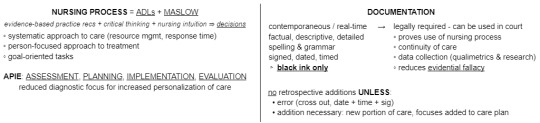
NATURE OF NURSING: no classes today but started the beginnings of some digital notes on intro nursing theory, here's what my unfinished sheet on ADLs looks like so far lol!!

HEALTH & APPL. BIOSCIENCES: CARDIOVASCULAR SYSTEM TODAY AUGHHHH i'm TELLING YOU they're releasing NEW BIOLOGY bro WTF IS THIS SHIT !!!!!
blood pressure: force of blood pushing against inside walls of vessels
heart position: slightly left of midline deep to the sternum in the mediastinum (compartment of thorax)
pericardium: tough sac restricts ♡ mvmt -> only moves slightly in thorax
fibrous p.c. = outer covering of tough dense connective tissue
serous p.c. = parietal layer (lines inner surface of fibrous p.c.) + visceral layer (epicardium, covers outer surface of ♡)
pericardial cavity = space between parietal & visceral layers
♡ walls = 3 distinct layers
epi = visceral layer of serous p.c. + connective tissue
myo = cardiac muscle
endo = lining of ♡ chamber (touches blood)
external ♡ anatomy: bottom point of ♡ = inferior apex; top point of ♡ = superior base -> 2x small superior atria + 2x larger inferior ventricles // separated by coronary sulcus (deep groove, lets veins & arts sit nicely on surface)
aneurysm: abnormal swelling of heart or artery/vein walls // varicose veins -> weakened valves
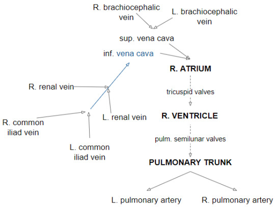
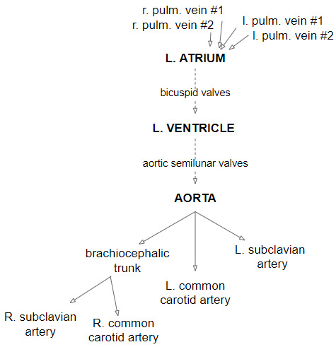
and now i present to you my desperate attempt to understand all these new tubes I JUST REALIZED I FORGOT THE CORONARY SINUS DRAINING INTO THE R. ATRIUM AUGHHHHH i also drew a diagram on paper of the heart yaaay
0 notes
Text
No, it's made of epicardium, myocardium, and endocardium.
@gotogrell come to my office.
17 notes
·
View notes
Photo
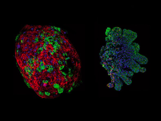
Bad Timing
Your heart isn't all muscle. Its muscle layer (myocardium) is sandwiched between endocardium and epicardium. Defects in the development of these layers can cause congenital heart disease (CHD), which affects one in 100 newborns. CHD can be caused by a coronary artery fistula (CAF) where a channel, partly comprising smooth muscle, develops between the coronary artery and another part of the heart. A defect in myocardium development may be to blame. Researchers investigated in chick embryos, damaging the myocardium before the epicardium layer forms. The result? Endocardium cells met epicardium cells too early, leading to CAF-like structures. The team then grew quail embryo endometrium (pictured, left) with chick embryo epicardium (right). Fluorescence microscopy revealed that where these different cells met, smooth muscle developed, and where they were apart, smooth muscle was absent. This supports the idea that CAFs form due to the inappropriately timed meeting of these cells.
Written by Lux Fatimathas
Image from work by P. Palmquist-Gomes and colleagues
Department of Animal Biology, Faculty of Sciences, University of Málaga, Málaga, Spain
Image originally published with a Creative Commons Attribution 4.0 International (CC BY 4.0)
Published in Experimental & Molecular Medicine, January 2023
You can also follow BPoD on Instagram, Twitter and Facebook
#science#biomedicine#immunofluorescence#congenital heart defects#myocardium#epicardium#endocardium#heart#heart muscle
4 notes
·
View notes
Photo


OMG stop!!
31 notes
·
View notes
Video
tumblr
Anatomy lesson
“The cardiovascular system is composed by the heart and by the blood vessels in which the blood flows continuously. The heart is the pump that gives the blood the thrust to flow inside the blood vessels. The blood vessels are like tubes of different diameters, used by the blood to reach cells and tissues; they can be divided into arteries that lead the blood from the heart to the rest of the body and into veins that bring back the blood to the heart. Between them the capillaries.
The heart is the central part of the whole system. Its contractions are the reason of the blood flow. It is placed close to the anterior wall of the thorax, right behind the sternum. It is inside the pericardial cavity, between the pulmonary pleurae, inside of the mediastinum. It is not perfectly centered, if we consider the body’s simmetry plan. It is for 2/3 of its volume in the left portion of the body and for 1/3 on the right portion. Its shape is a truncated cone with the base on the top and the apex on the botto,. It lies between the 3rd and the 6th intercostal space. It is an hollow organ composed of two atria and two ventricules. It can also be subdivided into the right and the left heart. between the atrium and its ventricule there’s a cusp valve; between the ventricle and the circulation there’s a semilunar valve. Valves prevent the reflux of the blood. The heart is wrapped by the pericardium divided into two layers: one is in contact with the surrounding organs, the other directly touches the heart. In between the the pericardial cavity filled by a lubricant fluit which prevents friction during the heart movements. The heart wall is divided into the epicardium, made by mesothelium and connective tissue; the myocardium, composed of heart muscles cells, vessels, connective tissue and nerves; the endocardium as a single layer of endothelial cells...”
Who gets the different references in the drawing wins an academic kiss.
107 notes
·
View notes
Text
Thing probably no one but me cares about but in that meta post I accidentally call the pericardium the epicardium which is kind of a calling a rectangle a square situation since the epicardium is the visceral layer of the pericardium and within the context of cardiac tamponade, it still makes sense and I just realized I'm pedantic-ing myself lol.
8 notes
·
View notes