#drbhishmachowdary
Explore tagged Tumblr posts
Visit Tumblr Blog
Explore Tumblr blogs with no restrictions, modern design and the best experience.
Last Seen Tumblr Blogs


Fun Fact
Celebrities use Tumblr as well.
Text
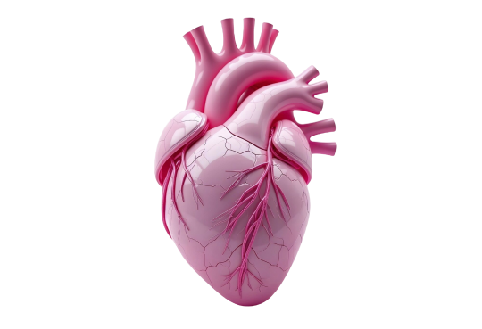
Valvular Heart Disease
#Valvular
#Heart
#Disease
#Medication
#Adults
#Transcatheter
#Aortic
#Pericutaneous
Use of medications (particularly HMG-CoA reductase inhibitors) to delay AS progression in asymptomatic patients has been studied and the results thus far are inconclusive.
Transcatheter techniques Balloon aortic valvuloplasty was introduced as a nonsurgical alternative treatment for AS and is integral to the management of congenital AS in the pediatric population.
Results in the adult population are not as good as anticipated because of procedural complications and high restenosis rates.
Aortic valvuloplasty is currently not an acceptable alternative to surgical AVR and its use should be restricted to palliative therapy in patients deemed nonoperable and as a bridge to AVR in hemodynamically unstable patients.
Percutaneous aortic valve replacement is currently being performed at experienced centers and may be a valuable treatment approach in the future. Current use is restricted to patients who are not surgical candidates.
https://drbhishmacardiologist.com/bicuspid-aortic-valve
#DrBhishmaChowdary
#BestCardiologistInHyderabad
#BestInterventionalCardiologistInHyderabad

#cardiology#health#healthcare#doctor#health tips#anatomical heart#health and wellness#heart doctor#cardiovascularhealth#heart specialist#drbhishma#drbhishmachowdary#drbhishmachowdarydonepudi#bestcardiologistinhyderabad
2 notes
·
View notes
Text
Mitral Valve Prolapse : Barlow's Syndrome , Floppy Mitral Valve Syndrome
Mitral Valve Prolapse
(Barlow’s syndrome, Floppy Mitral Valve Syndrome)
#MitralValveProlapse
#BarlowsSyndrome
#FloppyMitralValveSyndrome
#Degeneration
#Tissue
#Causes
#Abnormality
Causes
Primary or classic mitral valve prolapse (MVP) is an idiopathic valvular abnormality characterized by myxomatous degeneration of the mitral valve leaflets and subvalvular apparatus in the absence of any recognizable systemic connective tissue disorder.
It can be inherited as an autosomal dominant phenotype with incomplete penetrance.
Secondary MVP occurs in the setting of known connective tissue disorders, including Marfan’s syndrome, Ehlers–Danlos syndrome, pseudoxanthoma elasticum, and osteogenesis imperfecta.
MVP is also frequently associated with atrial septal defects and Ebstein’s anomaly.
One can also see acquired leaflet prolapse (i.e., functional MVP) in ischemic heart disease and dilated cardiomyopathy.
Overall prevalence is approximately 2% and is similar in men and women
#DrBhishmaChowdary
#BestCardiologistInHyderabad

#cardiology#health#healthcare#doctor#health tips#anatomical heart#health and wellness#heart doctor#heart specialist#cardiovascularhealth#drbhishma#drbhishmachowdary#bestcardiologistinhyderabad
0 notes
Text
Pathophysiology
Pathophysiology
#Pathophysiology
#LeftAtrium
#Diastole
#Hypertension
#Atrial
#Fibrillation
Elevated left atrium (LA) pressure is required to drive blood through the narrowed mitral valve orifice during diastole, leading to pulmonary venous hypertension and exertional dyspnea.
Secondary pulmonary arterial hypertension leads to right ventricular (RV) hypertrophy and failure.
LV systolic function is preserved but as filling is impaired, adequate cardiac output (CO) cannot always be maintained, especially with exercise and tachycardia.
Onset of atrial fibrillation (AF) is associated with abrupt clinical deterioration due to both the loss of atrial systole and the fast heart rate, which shortens the diastolic filling period.
Clinical features
• Dyspnea on exertion, orthopnea, paroxysmal nocturnal dyspnea.
• Acute pulmonary edema may be precipitated by uncontrolled AF, exercise, pulmonary infection, anesthesia, and pregnancy.
• AF increases the risk of thromboembolism. Systemic embolism occurs in 20%–30% and usually originates in the dilated LA and LA appendage.
• Fatigue is due to reduced cardiac output reserve.
• Hemoptysis can occur for a variety of reasons: alveolar capillary rupture (pink frothy pulmonary edema); bronchial vein rupture (larger hemorrhage); blood-stained sputum of chronic bronchitis; pulmonary infarction (low CO, immobile patient).
• Chest pain similar to angina may occur in patients with pulmonary hypertension and RV hypertrophy, even with normal coronaries.
• The enlarging LA may compress surrounding structures, producing hoarse voice (left recurrent laryngeal nerve compression—Ortner’s syndrome), dysphagia (esophageal compression), and left lung collapse (left main bronchus compression).
#DrBhishmaChowdary
#BestCardiologistInHyderabad

#cardiology#health#healthcare#doctor#health tips#anatomical heart#health and wellness#heart doctor#heart specialist#cardiovascularhealth#drbhishma#drbhishmachowdary#bestcardiologistinhyderabad
0 notes
Text
Aortic Dissection : Management
Aortic Dissection : Management
#AorticDissection
#Management
#Stabilization
#Treatment
#Diagnosis
Stabilize the patient
• If the diagnosis is suspected, transfer the patient to an area where full resuscitation facilities are readily available.
• Secure venous access with large-bore cannulas.
• Take blood for CBC, chemistries, and cross-match (10 units).
• When the diagnosis is confirmed or in cases with cardiovascular complications, transfer to ICU, insert an arterial line (radial unless the subclavian artery is compromised when a femoral line is preferred), central venous line, and urinary catheter.
• Immediate measures should be taken to correct blood pressure (see below).
• Give adequate analgesia (morphine 2.5–10 mg IV and metoclopramide 10 mg IV).
Plan definitive treatment
This depends on the type of dissection and its effects on the patient. General principles are as follows:
• Patients with involvement of the ascending aorta should have emergency surgical repair and BP control.
• Patients with dissection limited to the descending aorta are managed initially medically with aggressive blood pressure control.
However, this may change in the near future with emerging encouraging data from deployment of endovascular stent-grafts. Indications and principles for surgery
• Involvement of the ascending aorta
• External rupture (hemopericardium, hemothorax, effusions)
��� Arterial compromise (limb ischemia, renal failure, stroke)
• Contraindications to medical therapy (AR, CHF)
• Progression (continued pain, expansion of hematoma on further imaging, loss of pulses, pericardial rub, or aortic insufficiency)
The aim of surgical therapy is to replace the ascending aorta, thereby preventing retrograde dissection and cardiac tamponade (the main cause of death). The aortic valve may need reconstruction and resuspension unless it is structurally abnormal (bicuspid or Marfan’s), when it is replaced.
#DrBhishmaChowdary
#BestCardiologistInHyderabad
#DrBhishmaChowdary
#BestCardiologistInHyderabad

#cardiology#health#healthcare#doctor#health tips#anatomical heart#health and wellness#heart doctor#heart specialist#cardiovascularhealth#drbhishma#drbhishmachowdary#bestcardiologistinhyderabad
0 notes
Text
Hypertensive Emergencies : Management
Hypertensive Emergencies : Management
#Hypertension
#Emergencies
#Diagnosis
#Priorities
#LongTermTreatment
Priorities in management are as follows :
1. Confirm the diagnosis and assess the severity.
2. Identify those patients needing specifi c emergency treatment.
3. Plan long-term treatment.
Diagnosis and severity
• Ask about previous BP recordings, previous and current treatment, sympathomimetics, antidepressants, nonprescription drugs, and recreational drugs.
• Check the blood pressure yourself, in both arms, after a period of rest and, if possible, on standing.
• Examine carefully for clinical evidence of cardiac enlargement or heart failure, peripheral pulses, renal masses, or focal neurological deficit.
Always examine the fundi—dilate if necessary.
All patients should have the following tests:
• CBC Microangiopathic hemolytic anemia with malignant hypertension • Chemistries Renal impairment and/or dK+ (diffuse intrarenal ischemia and secondary hyperaldosteronism)
• Coagulation screen Disseminated intravascular coagulation (DIC) with malignant hypertension
• CXR Cardiac enlargement Aortic contour (dissection?) Pulmonary edema
• Urinalysis Protein and red cells ± casts Other investigations depending on clinical picture and possible etiology include the following:
• 24-hour urine collection Creatinine clearance Free catecholamines, metanephrines or vanilmandellic acid (VMA)
• ECHO LVH, aortic dissection
• Renal US and Doppler Size of kidneys and renal artery stenosis
• MR renal angiogram Renal artery stenosis
• CT/MR brain Intracranial bleed
• Drug screen Cocaine, amphetamine, others Indications for admission
• Diastolic blood pressure persistently t120 mmHg
• Retinal hemorrhages, exudates or papilledema
• Renal impairment
https://drbhishmacardiologist.com/systemic-hypertension
#DrBhishmaChowdary
#BestCardiologistInHyderabad

#cardiology#health#healthcare#doctor#health tips#anatomical heart#health and wellness#heart doctor#heart specialist#cardiovascularhealth#drbhishma#drbhishmachowdary#bestcardiologistinhyderabad
0 notes
Text
Birth Control Pills : Heart Attack
Birth Control Pills : Heart Attack
#Heart
#Birth
#Control
#Pills
#HeartAttack
Studies show that women who use high-dose birth control pills (oral contraceptives) are more likely to have a heart attack or stroke because blood clots are more likely to form in the blood vessels.
These risks are lessened once the birth control pill is stopped. Using the pill also may worsen the effects of other risk factors, such as smoking, high blood pressure, diabetes, high blood cholesterol, and overweight.
Much of this information comes from studies of birth control pills containing higher doses of hormones than those commonly used today. Still, the risks of using low-dose pills are not fully known.
Therefore, if you are now taking any kind of birth control pill or are considering using one, keep these guidelines in mind:
Don’t mix smoking and the “pill.”
If you smoke cigarettes, stop smoking or choose a different form of birth control. Cigarette smoking raises the risk of serious health problems from birth control pill use, especially the risk of blood clots.
For women over 35, the risk is particularly high. Women who use birth control pills should not smoke. Pay attention to diabetes.
Levels of glucose, or blood sugar, sometimes change dramatically in women who take birth control pills.
If you are diabetic or have a close relative who is, be sure to have regular blood sugar tests if you take birth control pills. Watch your blood pressure.
After starting to take birth control pills, your blood pressure may go up. If your blood pressure increases to 140/90 mmHg or higher, ask your doctor about changing pills or switching to another form of birth control.
Be sure to get your blood pressure checked at least once a year. Talk with your doctor. If you have heart disease or another heart problem, or if you have suffered a stroke, birth control pills may not be a safe choice.
Be sure your doctor knows about these and any other serious health conditions before prescribing birth control pills for you.
#DrBhishmaChowdary
#BestCardiologistInHyderabad
#cardiology#health#healthcare#doctor#health tips#anatomical heart#health and wellness#heart doctor#heart specialist#cardiovascularhealth#drbhishma#drbhishmachowdary#bestcardiologistinhyderabad
0 notes
Text
New Risk Factors ? Healthy Heart
New Risk Factors? Healthy Heart
#New ❤️
#RiskFactors 🤎
#Health 💛
#Heart 🩷
#Treatment 💚
#Cholesterol ����
#BloodPressure 🧡
We know that major risk factors such as high blood cholesterol, high blood pressure, and smoking boost heart disease risk. Researchers are studying other factors that might contribute to heart disease, including inflammation of the artery walls. Several emerging risk factors have been identified.
We don’t know for sure yet whether they lead to heart disease or whether treating them will reduce risk. While these possible risk factors are not recommended for routine testing, ask your doctor whether you should be tested for any of them. C-reactive protein (CRP).
High levels of CRP may indicate inflammation in the artery walls. A simple blood test can measure the levels of CRP in the blood. In many cases, a high CRP level is a sign of metabolic syndrome.
Treatment of the syndrome with lifestyle changes—weight loss and regular physical activity—can often lower CRP. Homocysteine. High blood levels of this amino acid may increase risk for heart disease.
It may be possible to lower elevated levels of homocysteine by getting plenty of folic acid and vitamins B6 and B12 in your diet. Lp(a) protein. This lipoprotein may make it easier for blood clots to form. Niacin, a cholesterol-lowering drug, may help to lower Lp(a) protein levels.
#DrBhishmaChowdary 🩺
#BestCardiologistInHyderabad 🥼
https://drbhishmacardiologist.com/heart-disease-treatment
#cardiology#health#healthcare#doctor#health tips#health and wellness#anatomical heart#heart doctor#heart specialist#cardiovascularhealth#drbhishmachowdary#bestcardiologistinhyderabad
0 notes
Text
Mineral Medicine
Mineral Medicine
Mineral Medicine :
#Minerals 💛
#Control 💜
#BloodPressure ❤️
#Heart 🩵
#Health 💚
Another Way To Control Blood Pressure Certain mineral-rich foods can help keep blood pressure levels healthy.
For example, a diet rich in potassium can help to both prevent and control high blood pressure. A potassium-rich diet not only blunts the effects of salt on blood pressure, but may also reduce the risk of developing kidney stones, and possibly decrease bone loss with age.
But be sure to get your potassium from food sources, not from supplements. Many fruits and vegetables, some dairy foods, and fish are rich sources of potassium. Calcium and magnesium are two other minerals that may help to prevent high blood pressure, as well as improve health in other ways.
Low-fat or fat-free milk and milk products are rich sources of calcium, while magnesium is found in many whole-grain products; dark green, leafy vegetables; fish; and dry beans.
https://drbhishmacardiologist.com/blogs
#DrBhishmaChowdary 🩺
#BestCardiologistInHyderabad 🥼

#cardiology#health#healthcare#doctor#health tips#anatomical heart#health and wellness#heart doctor#heart specialist#cardiovascularhealth#DrBhishmaChowdary#BestCardiologistInHyderabad
0 notes
Text
Aortic Stenosis
Aortic Stenosis
#AorticStenosis
#Valve
#Elderly
#Patients
#Helsinki
#Study
Calcific or degenerative aortic valve disease is the most common valvular abnormality seen in the elderly. The prevalence of at least moderate aortic stenosis (AVA <1.2 cm2 ) in patients aged 75–85 years old was 5% in the Helsinki Aging Study.
#DrBhishmaChowdary
#BestCardiologistInHyderabad

#cardiology#health#healthcare#doctor#health tips#anatomical heart#health and wellness#heart doctor#heart specialist#cardiovascularhealth#drbhishmachowdarydonepudi#drbhishma#bestcardiologistinhyderabad
0 notes
Text
Aortic Regurgitation
Aortic Regurgitation
#Aortic
#Regurgitation
#Valve
#Surgery
#Mortality
Aortic regurgitation is less common in the elderly than calcific aortic stenosis or mitral regurgitation and is most often associated with aortic stenosis or dilation of the ascending aorta from long-standing hypertension.
Although prophylactic aortic valve surgery is usually recommended for asymptomatic patients with severe aortic insufficiency (AI) and evidence of LV dysfunction, in the elderly (especially patients over the age of 80), it is recommended that aortic valve surgery be reserved for those patients with symptoms and severe AI, given the increased risk of operative and long-term mortality with increasing age
#DrBhishmaChowdary
#BestCardiologistInHyderabad

#cardiology#health#healthcare#doctor#health tips#anatomical heart#health and wellness#heart doctor#heart specialist#cardiovascularhealth#drbhishmachowdarydonepudi#drbhishma#bestcardiologistinhyderabad
0 notes
Text
Mitral Stenosis
Mitral Stenosis
#Mitral
#Stenosis
#Patients
#Younger
#Calcification
#Replacement
Mitral stenosis remains a disease primarily of younger patients, with rheumatic heart disease being the most common etiology. In elderly patients the most common cause of mitral stenosis is impingement on the mitral valve by mitral annular calcification.
The preferred surgical treatment for mitral stenosis is mitral commissurotomy. However, this is often not possible in the elderly, necessitating mitral valve replacement. The ideal treatment for those elderly patients with favorable valvular morphology is percutaneous valvotomy. However, the number of elderly patients who have valvular morphology amenable to this technique is quite limited.
#DrBhishmaChowdary
#BestCardiologistInHyderabad

#cardiology#health#healthcare#doctor#health tips#anatomical heart#health and wellness#heart doctor#heart specialist#cardiovascularhealth#drbhishmachowdarydonepudi#drbhishma#bestcardiologistinhyderabad
0 notes
Text
Mitral Regurgitation
Mitral Regurgitation
#Mitral
#Regurgitation
#Causes
#Acute
#Chronic
#Dysfunction
#Trauma
Causes
Acute
• Infective endocarditis
• Acute myocardial infarction (MI) (usually inferior wall) from papillary muscle dysfunction or acute rupture
• Trauma
Chronic
• Mitral valve prolapse
• Rheumatic heart disease
• Ischemic heart disease
• Left ventricular dilatation of any cause
• Hypertrophic cardiomyopathy
• Carcinoid syndrome
• Fen-phen valvulopathy
• Congenital lesions (i.e., cleft mitral valve).
#DrBhishmaChowdary
#BestCardiologistInHyderabad

#cardiology#health#healthcare#doctor#health tips#anatomical heart#health and wellness#heart doctor#heart specialist#cardiovascularhealth#drbhishmachowdarydonepudi#drbhishma#bestcardiologistinhyderabad
0 notes
Text
Aortic Root Disease
#Aortic
#Root
#Disease
#Dilation
#Hypertension
#Syphilis
#Biscuspid
#Valve
Dilatation, aneurysm, and dissection cause failure of coaptation of cusps. One may see diastolic cusp prolapse in dissection.
Causes
• Hypertension
• Marfan’s syndrome
• Osteogenesis imperfecta
• Syphilis
• Spondyloarthritides (ankylosing spondylitis, Reiter’s, etc.)
• Trauma
• Bicuspid aortic valve
https://drbhishmacardiologist.com/bicuspid-aortic-valve
#DrBhishmaChowdary
#BestCardiologistInHyderabad

#cardiology#health#healthcare#doctor#health tips#anatomical heart#health and wellness#heart doctor#heart specialist#cardiovascularhealth#drbhishmachowdarydonepudi#drbhishma#bestcardiologistinhyderabad
0 notes
Text
Valve Hemodynamics
Valve Hemodynamics
#Valve
#Hemodynamics
#Dimension
#OrificeArea
#Homografts
Different prosthetic valves have unique profiles and valve areas. For any given valve dimension, bioprosthetic valves and ball and cage valves have the smallest effective valve orifice area, and homografts have the largest valve area (comparable to native valve area).
https://drbhishmacardiologist.com/bicuspid-aortic-valve
#DrBhishmaChowdary
#BestCardiologistInHyderabad

#cardiology#health#healthcare#doctor#health tips#anatomical heart#health and wellness#heart doctor#heart specialist#cardiovascularhealth#drbhishmachowdarydonepudi#drbhishma#bestcardiologistinhyderabad
0 notes
Text
Hemolysis
Hemolysis
#Hemolysis
#Prostheses
#Investigation
#Treatment
#Valve
#Transfusion
#FolicAcid
A low level of background hemolysis is common in patients with mechanical prostheses (even when functioning normally). Severe hemolysis is uncommon and is usually secondary to valve dysfunction (paravalvular leak, dehiscence, infection). Hemolysis is uncommon in tissue prostheses.
• Investigation: anemia, elevated LDH, low serum haptoglobin level, reticulocytosis, schistocytes on peripheral smear.
• Treatment: treat underlying problem (including further valve surgery if needed); give blood transfusion, folic acid, and iron supplementation
https://drbhishmacardiologist.com/aortic-stenosis
#DrBhishmaChowdary
#BestCardiologistInHyderabad

#cardiology#health#healthcare#doctor#health tips#anatomical heart#health and wellness#heart doctor#heart specialist#cardiovascularhealth#drbhishma#drbhishmachowdarydonepudi#bestcardiologistinhyderabad
0 notes
Text
Echocardiography
Echocardiography
#Echocardiography
#Sensitivity
#Diognosis
#Imaging
#Clinical
#Suspicion
TTE has a low sensitivity of <50% but a high specificity of 98% for the diagnosis of infective endocarditis. TEE has a sensitivity of 75%–95% and a specificity of 85%–98% and is the recommended diagnostic imaging test in patients with prosthetic valves, cases rated at least possible IE by clinical criteria, and patients with complications. TEE is also recommended when the clinical suspicion for IE is high despite a negative TTE.
https://drbhishmacardiologist.com/advanced-coronary-physiology
#DrBhishmaChowdary
#BestCardiologistInHyderabad

#cardiology#health#healthcare#doctor#health tips#anatomical heart#health and wellness#heart doctor#heart specialist#cardiovascularhealth#drbhishma#drbhishmachowdarydonepudi#bestcardiologistinhyderabad
0 notes