#distantinfluence
Text
СИСТЕМА БЕСКОНТАКТНОЙ РЕГИСТРАЦИИ РЕАКЦИИ
СИСТЕМА БЕСКОНТАКТНОЙ РЕГИСТРАЦИИ РЕАКЦИИ ЧЕЛОВЕКА-ОПЕРАТОРА И ГРУППЫ ЛЮДЕЙ НА ИНФОРМАЦИОННО-ПСИХОЛОГИЧЕСКИЕ ВОЗДЕЙСТВИЯ
Орлов Д.В.1,2, Коротков К.Г. 1,2, Гатчин Ю.А. 1, Сухостат В.В. 1, Гришенцев А.Ю. 1
1 Санкт-Петербургский национальный исследовательский университет информационных технологий, механики и оптики
2 Федеральное государственное бюджетное учреждение «Санкт-Петербургский научно- исследовательский институт физической культуры»,
Метод газоразрядной визуализации (ГРВ) используется во множестве научных и практических областей . Одним из новых направлений является регистрация реакции группы людей на различные информационно-психологические воздействия . Для проведения таких измерений авторами совместно с компанией ООО "КТИ" были разработаны и запущены в серийное производство прибор «ГРВ Эко-Тестер» и антенна «ГРВ Спутник», которые обеспечивают необходимую чувствительность и стабильность измерительной системы. Прибор «ГРВ Эко-Тестер» был спроектирован в соответствии с рекомендациями по анализу и коррекции дестабилизирующих факторов в процессе измерений методом ГРВ . Была разработана стандартная процедура проведения измерений и обработки результатов .
В случае регистрации реакции человека-оператора или групп операторов на различные виды информационно-психологических воздействий возможна реализация двух подходов:
1) мониторинг состояния окружающего операторов пространства по всевозможным характеристикам с целью своевременного выявления факторов, способных оказать влияние на функциональное состояние человека;
2) мониторинг функционального состояния самих операторов.
В случае осуществления первого подхода необходима установка множества датчиков, регистрирующих уровни шумового давления, вибрации, инфразвука, ультразвука, электромагнитных полей промышленных частот, электростатических полей, магнитных полей, радиации, микроклимата, концентрации легких аэроионов, химический состав воздуха. Кроме этого необходимо специальное программное обеспечение, позволяющее централизованно контролировать сигналы от всех вышеперечисленных датчиков и приборов. С технической и коммерческой точек зрения данная задача является нетривиальной.
В случае же реализации второго подхода обычно проводится измерение различных физиологических характеристик (вариабельность сердечного ритма, артериальное давление, проводимость кожи, движения глаз, выражение лица, виброизображение) каждого человека-оператора в реальном времени. В подобном случае кроме сложности осуществления подобной задачи (особенно в случае бесконтактной регистрации) добавляется некомфортность для человека-оператора наличия множества датчиков на теле (контактная регистрация), что сказывается на продуктивности и эффективности работы. Вариантом решения данной проблемы является использование бесконтактных датчиков и технологий, позволяющих неинвазивно получать информацию о функциональном состоянии человека. Однако на данный момент разработаны системы, позволяющие определять функциональную психофизиологическую активность лишь отдельного человека. Следовательно, для контроля состояния группы людей требуется большое количество бесконтактных систем, кратное количеству человек в группе, что означает большие финансовые затраты, хотя и даёт возможность отслеживать состояние каждого члена группы в отдельности.
Принимая во внимание трудности реализации традиционных подходов, возникает потребность в новых методах, позволяющих осуществлять неселективный мониторинг характеристик окружающей среды и неинвазивную оценку функционального состояния группы людей в реальном времени. Таким подходом является метод газоразрядной визуализации, а именно прибор «ГРВ Эко-Тестер» с антенным датчиком «ГРВ Спутник». Целью данной работы является экспериментальное тестирование прибора «ГРВ Эко- Тестер» для определения адекватности получаемых данных поставленным задачам: возможность выявления физических и химических воздействий, влияющих на состояние оператора, и точность реакции на изменение функционального состояния человека- оператора.
Методы исследований
Принцип формирования газоразрядный изображений
Принцип формирования газоразрядных изображений (ГРИ) описан в .
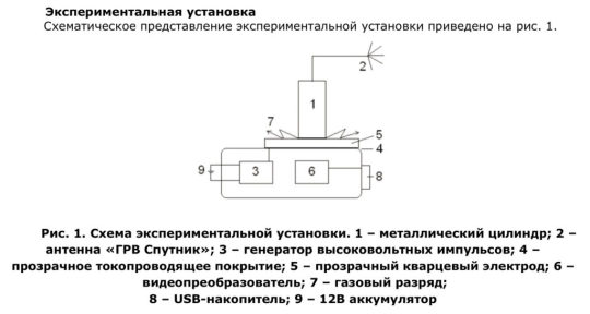
Процедура формирования ГРИ с помощью прибора «ГРВ Эко-Тестер» заключается в следующем. Металлический цилиндр (тест-объект) помещается на прозрачный кварцевый электрод, на обратную сторону которого нанесено прозрачное токопроводящее покрытие, на которое в течение заданного промежутка времени подаются импульсы напряжения от генератора. Мощность импульсов и длительность воздействия задаются программно на персональном компьютере. При высокой разности потенциалов между тест-объектом и пластиной из металлического цилиндра выбиваются электроны и фотоны, которые, сталкиваясь с молекулами воздуха, ионизируют их, что в итоге приводит к развитию лавинного и/или скользящего газового разряда. Характеристики газового разряда определяются свойствами внешней цепи – то есть тест-объекта, провода, подключенного к нему, антенны «ГРВ Спутник» и пространства между антенной и землей. Пространственное распределение разряда фиксируется специализированной видеокамерой на базе ПЗС-матрицы, расположенной непосредственно под прозрачным электродом.
Видеопреобразователь осуществляет оцифровку изображения и передачу его на компьютер для дальнейшей обработки. ГРИ обрабатываются в специально разработанном программном комплексе, где осуществляется расчет параметров изображений, таких как энергия свечения, площадь засветки, средняя интенсивность разряда и др. Параметры ГРИ зависят от физических характеристик внешней цепи, в частности, электрической емкости и сопротивления .
GDV Sputnik distant influence
Также существует техническая возможность обработки ГРИ в реальном времени на сервере, на котором установлено специально разработанное программное обеспечение BioDeck. Прибор ГРВ через Интернет посылает каждое снятое ГРИ на сервер, где рассчитываются необходимые параметры. Рассчитанные данные в виде графиков пересылаются с сервера на пользовательский компьютер, на котором установлена пользовательская версия программного обеспечения BioDeck, позволяющая получать числовые значения рассчитанных параметров ГРИ в реальном времени.
GDV Sputnik distant influence
Full text, PDF: GDV Sputnik distant influence
Read the full article
#Bio-WellSputnik#Bio-WellSputnikSensor#distant#distantinfluence#GDVSputnik#GDVSputnikdistantinfluence#ГРВСпутник
0 notes
Text
Music waves Research and the Biointernet
Music waves Research and the Biointernet
https://www.iumab.org/category/music/
When a musician is playing a piece, and the audience is enjoying it, they can develop physical synchronies.
The team used a technique called near-infrared spectroscopy to monitor the brain activity of a professional violinist while he was videoed playing a series of 12 brief, classical pieces. They then used the same technique (which involves shining beams of light through the skull, to monitor changes in blood flow) on 16 women while they watched the video, and listened to all of these pieces. (Because gender differences in inter-brain synchronisation have previously been observed, only women were recruited as listeners.)

BIOELECTROGRAPHY OF MUSICAL ENVIRONMENT
The averaged inter-brain coherence between the audience and a violinist predicts the popularity of violin performance
Highlights
Music popularity is explored in terms of interpersonal brain synchronization.•
Dual-NIRS approach is used to record brain activity of the violinist and audience.•
The averaged IBC between audiences and the violinist correlates with popularity.•
IBC not only discriminates high and low popularity, but also predicts popularity.•
Music appreciation involves brains in a temporally aligned network.
Abstract
Why is some music well-received whereas other music is not? Previous research has indicated the close temporal dependencies of neural activity among performers and among audiences. However, it is unknown whether similar neural contingencies exist between performers and audiences. Here, we used dual near-infrared spectroscopy (NIRS) to assess whether inter-brain synchronization between violinist and audience underlies the popularity of violin performance. In the experiment, individual audience members (16 females) watched pre-recorded videos, each lasting 100 s or so, in which a violinist performed 12 musical pieces. The results showed that the popularity of the performance correlated with the left-temporal inter-brain coherence (IBC) between the audience and the violinist. The correlation was stronger at late watching (>50 s) than at early watching (≤50 s). The smaller the Granger causality from the audience to the violinist was, the higher was the popularity of the piece with the audience. Discriminant analysis showed that the IBC could distinguish high popularity from low popularity. Further analysis using support vector regression showed that the IBC could also predict the popularity. These findings reveal the association of IBC with the popularity of violin performance. Music appreciation involves the brains of music producers and perceivers in a temporally aligned network through which audiences perceive the intentions of the performer and show positive emotions related to the musical performance.
IBC, Performance popularity, Violin, Temporal, cortex, NIRS
https://www.sciencedirect.com/science/article/pii/S1053811920301427?dgcid=rss_sd_all
https://digest.bps.org.uk/2020/03/23/musicians-and-their-audiences-show-synchronised-patterns-of-brain-activity/
https://www.iumab.org/category/music/
IUMAB Music Research Library
Music Waves, GDV Sputnik, Brain activity and more
Welcome! Music waves Research and the Biointernet
See also:
2020 Influence of mobile phone to people
2020 Influence of mobile phone to people
IUMAB Music Research Library
Influence of mobile phone to the brain
Response mobile phone to people
2020 Music structuration of water
2020 Music structuration of water
IUMAB Music Research Library
Bioelectrography Water Research
2020 Remote detection of music influence
2020 Remote detection of music influence
IUMAB Music Research Library
Distant influence Research
Read the full article
#cortex#distantinfluence#IBC#mobilephoneandhealth#mobilephoneradiations#Music#musicResearch#NIRS#Non-localConsciousness#nonlocal#nonlocalinteraction#Performancepopularity#Remotedetectionofmusicinfluence#Temporal#Violin#Water
0 notes
Text
Optical communication channels in the brain
Are there optical communication channels in the brain?
Parisa Zarkeshian, Sourabh Kumar, Jack Tuszynski, Paul Barclay, Christoph Simon (Submitted on 23 Aug 2017)
Despite great progress in neuroscience, there are still fundamental unanswered questions about the brain, including the origin of subjective experience and consciousness. Some answers might rely on new physical mechanisms. Given that biophotons have been discovered in the brain, it is interesting to explore if neurons use photonic communication in addition to the well-studied electro-chemical signals. Such photonic communication in the brain would require waveguides. Here we review recent work suggesting that myelinated axons could serve as photonic waveguides. The light transmission in the myelinated axon was modeled, taking into account its realistic imperfections, and experiments were proposed both in-vivo and in-vitro to test this hypothesis. Potential implications for quantum biology are discussed.
Comments:13 pages, 5 figures, review of arXiv:1607.02969 for Frontiers in Bioscience, updated figures, new references on existence of opsins in the brain and experimental effects of light on neuronsSubjects:Biological Physics (physics.bio-ph); Optics (physics.optics); Neurons and Cognition (q-bio.NC); Quantum Physics (quant-ph)Cite as:arXiv:1708.08887 (or arXiv:1708.08887v1 for this version)
Submission history
From: Parisa Zarkeshian
Wed, 23 Aug 2017 22:54:52 UTC (1,981 KB)
1. ABSTRACT
Despite great progress in neuroscience, there are still fundamental unanswered questions about the brain, including the origin of subjective experience and consciousness. Some answers might rely on new physical mechanisms. Given that biophotons have been discovered in the brain, it is interesting to explore if neurons use photonic communication in addition to the well-studied electro-chemical signals. Such photonic communication in the brain would require waveguides. Here we review recent work suggesting that myelinated axons could serve as photonic waveguides. The light transmission in the myelinated axon was modeled, taking into account its realistic imperfections, and experiments were proposed both in vivo and in vitro to test this hypothesis. Potential implications for quantum biology are discussed.
2. INTRODUCTION
Over the past decades a substantial number of facts has been discovered in the field of brain research. However, the fundamental question of how neurons, or more specifically all particles involved in the biological processes in the brain, contribute to mental abilities such as consciousness is still unanswered. The true explanation to this question might rely on physical processes other than those that have been discovered so far. One interesting candidate to focus on is biophotons, which might serve as supplementary information carriers in the brain in addition to the well established electro-chemical signals.
Biophotons – which are photons ranging from near-IR to near-UV frequency and emitted without any enhancement or excitation– have been observed in many organisms such as bacteria (1), fungi (2), germinating seeds (3), plants (4), animal tissue cultures (5), and different parts of the human body (6–9), including the brain (10–15). These biophotons are produced by the decay of electronically excited species which are created chemically during oxidative metabolic processes (16, 17) and can contribute to communication between cells (18). Moreover, several experimental studies show the effects of light on neurons’ and, generally, the brain’s function (19–21). The existence of biophotons and their possible effects on the the brain along with the fact that photons are convenient carriers of information raises the question whether there could be optical communication in the brain.
For the sources and detectors of the optical communication process in the brain, mitochondrial respiration (22, 23) or lipid oxidation (24), and centrosomes (25) or chromophores in the mitochondria (26) have been proposed, respectively. It has also been observed that opsins, photoreceptor protein molecules, exist in the brains of birds (27, 28), mammals (29–32), and more general vertebrates (33) and even in other parts of their bodies (34, 35) as well.
Another essential element for this optical communication, which is not well established yet, is the existence of physical links to connect all of these spatially separated agents in a selective way. In the dense and (seemingly) disordered environment of the brain, waveguide channels for traveling photons would be the only viable way to achieve the targeted optical communication processes. Mitochondria and microtubules in neurons have been introduced as the candidates for such waveguides (36–39). How- ever, they are not suitable in reality due to their small and inhomogeneous structure for light guidance over proper distances in the brain.
Ref. (40) proposed myelinated axons as potential biophoton waveguides in the brain. The proposal is supported by a theoretical model and numerical results taking into account real imperfections. Myelin sheath (formed in the central nervous system by a kind of glia cell called oligodendrocyte) is a lamellar structure surrounding the axon and has a higher refractive index (41) than both the inside of the axon and the interstitial fluid outside (see Fig. 1a) which let the myelin sheath to guide the light inside itself for optical communications. This compact sheath also increases the propagation speed of an action potential (via saltatory conduction) based on its insulating property (42). There has been a few indirect experimental evidence for light conduction by axons (12, 43, 44). Another related and interesting experiment has shown that a certain type of glia cells, known as Mu ̈ller cells, guide light in mammalian eyes (45, 46). Ref. (40) also proposed experiments to test the existence of the optical waveguides in the brain.
One interesting property of optical communication channels is that they can also transmit quantum information. Quan- tum effects in biological systems are being studied in different areas such as photosynthesis (47, 48), avian magnetoreception (49, 50), and olfaction (51, 52). There is an increasing number of conjectures about the role of quintessential quantum features such as superposition and entanglement (53) in the brain (15, 38, 54–56). The greatest challenge when considering quantum effects in the brain or any biological system in general is environmentally induced decoherence (57), which leads to the suppression of these quantum phenomena. However, some biological processes can be fast and may show quantum features before they are destroyed by the environment. Moreover, nuclear spins can have coherence times of tens of milliseconds in the brain (58, 59). A recent proposal on “quantum cognition” suggests even longer coherence times of nuclear spins (56), but relies on quantum information transmission via molecule transport, which is very slow. In contrast, photons are the fastest and most robust carriers for quantum information over long distances, which is why currently man-made quantum networks rely on optical communication channels (typically optical fibers) between spins (60, 61).
3. Results
To show that myelinated axons could serve as the waveguides for traveling biophotons in the brain, Ref. (40) solved the three dimensional electromagnetic field equations numerically in different conditions, using Lumerical’s software packages FDTD (Finite Difference Time Domain) Solutions and MODE Solutions. These software packages solve Maxwell’s equations numerically, allowing the optical properties of dielectric structures defined over a mesh with subwavelength resolution to be simulated.
The refractive indices of the fluid outside of the axon, the axon, and the myelin sheath were taken close to 1.34, 1.38 and 1.44 respectively (see Fig. 1a), which are consistent with their typical values (41, 62, 63). These indexes let the myelin sheath guide the light inside itself. The ratio of the radius of the axon, r to the outer radius of the myelin sheath r′ (g-ratio) is taken equal to 0.6 for the most of the simulations, close to the experimental values (64). In reality, the radius of the myelinated axons in the brain changes from 0.2 microns to close to 10 microns (65). For the purpose of guiding light inside the myelin sheath, Ref. (40) considered the wavelength of the observed biophotons in the brain which is from 200 nm to 1300 nm. Since several proteins in the environment of the axons strongly absorb at wavelengths close to 300nm, a wavelength range of the transmitted light from the shortest permissible wavelength, λmin = 400nm, to the longest one, λmax, was chosen to avoid the absorption and confine the light well in the myelin sheath. λmax is chosen to the upper bound of the observed biophoton wavelength (1300 nm) or the thickness of the myelin sheath (denoted by d), whichever is smaller. Besides λmin and λmax, an intermediate wavelength was considered, denoted by λint , corresponding to the central permissible frequency (mid-frequency of the permissible frequency range) in the simulations.
In the following section we discuss the guided modes in the myelinated axons and their transmissions in nodal and para- nodal regions and even in the presence of the imperfections such as bends, varying cross-sections, and non-circular cross-sections.

Optical communication channels in the brain
Optical communication channels in the brain
See also: Consciousness Research, Human Light System, Biophotons
More Bioelectrography Research
Read the full article
#Biophotons#channelsinthebrain#Consciousness#ConsciousnessResearch#ConsciousnessStudies#distantinfluence#HumanLightSystem#Opticalcommunicationchannels#Opticalcommunicationchannelsinthebrain
0 notes
Last Seen Blogs

fabspin1
Untitled

est-guide
Гид в Таллинне

espinosatk353-blog
Esplendido website f-k 4-8

zombie-yokai
Requests are closed!

chandrasetiawaan-blog
chandra setiawan