#colour doppler scan
Explore tagged Tumblr posts
Visit Tumblr Blog
Explore Tumblr blogs with no restrictions, modern design and the best experience.
Last Seen Tumblr Blogs


Fun Fact
US Tumblr user growth rate is estimated to slow down to 4.1%.
Text

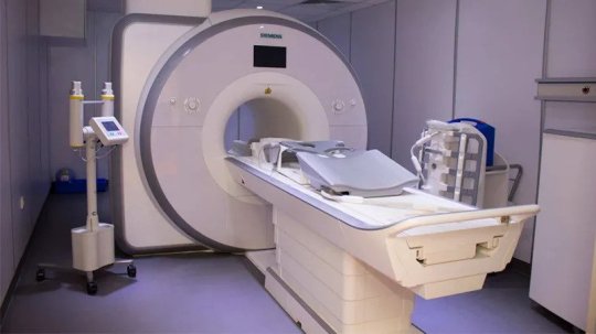
Need a trusted diagnostic center that puts your health first? At Kior Healthcare, we deliver top-quality medical imaging for accurate diagnoses and your peace of mind.
Why choose us?
Advanced Technology for Precise Results
Skilled & Caring Professionals
Patient-First Approach
Easy & Hassle-Free Access
Proudly Serving the Community
Experience modern diagnostics in a warm, patient-friendly environment at Kior Healthcare!
Visit our website: www.kiorhealthcare.com
Contact us: +91 8263000004
#diagnosticcentre #diagnosticcentreinmohali
#diagnostic centre#diagnostic services#diagnostic centre in mohali#top diagnostic centre in mohali#CT scan centre in mohali#Colour Doppler test in Mohali#MRI Scanning Centre in Mohali#Digital Mammography Test in Chandigarh
0 notes
Text
#pathlab#health#MRI#CT SCAN#Pathology#X RAY#BMD Dexa Scan#Colour Doppler & Ultrasound#3T MRI#Cardiology Test#Mammography#Neuroimaging#CBCT & Dental Imaging#Uroflowmetry
0 notes
Text
Ultrasound Innovation Progression: Changing Clinical Imaging With Sonograph
The field of clinical imaging is going through an interesting change, and at the cutting edge of this upset stands ultrasound innovation. With always advancing capacities and expanding moderateness, ultrasound is reshaping pre-birth care, especially in Mumbai, where focuses like NM Eva offer pregnancy ultrasound at open costs. This blog digs into the headways in sonography in Mumbai, eaturing its advantages for pregnant ladies and investigating the urgent job of colour doppler ultrasound in guaranteeing solid pregnancies.

Opening the Force of Sound Waves
Ultrasound, or sonography, uses sound waves to make ongoing pictures of inside organs and tissues. Its harmless nature and absence of radiation pursue it a favored decision for pregnant ladies, offering significant experiences into the fostering child's wellbeing.
A Brief look into Your Developing Marvel
Pregnancy ultrasound assumes an essential part in pre-birth care, giving key data that guides vital choices. From laying out your due date and affirming the quantity of embryos to checking fetal development and distinguishing expected peculiarities, these sweeps offer a window into your developing wonder.
NM Eva: Your Believed Sonography Accomplice in Mumbai
Situated in Mumbai, NM Eva is an all-ladies' demonstrative community eminent for its pregnancy sonography skill and reasonableness. Furnished with state of the art ultrasound innovation in Mumbai, we offer a scope of ultrasound administrations, including:
NT Scan: Directed in the first trimester, this sweep surveys the nuchal clarity, a liquid occupied space at the rear of the child's neck, to distinguish likely chromosomal irregularities.
Variety Doppler Ultrasound: This exceptional strategy utilizes sound waves and variety coded pictures to imagine blood stream in the placenta and umbilical rope, helping with the evaluation of fetal prosperity.
Deciphering the Blood Stream Story
Variety Doppler ultrasound merits unique notice for its crucial job in guaranteeing solid pregnancies. This innovation goes past essentially recording the child's pulse; it gives a point by point image of blood stream designs all through the baby, uncovering potential issues like development limitations or placental inadequacy.
NM Eva: Making Progressed Care Available
At NM Eva, understanding the significance of ideal conclusion in high-risk pregnancies, we focus on variety Doppler examines during the beginning phases. Nonetheless, for most pregnancies, these sweeps are commonly acted in the third trimester, ideally towards the end, to preclude any late-stage difficulties.
The Eventual fate of Sonography: More splendid, More clear, and Closer
With continuous headways in ultrasound innovation, what's in store holds energizing prospects. Anticipate much more honed pictures, expanded transportability, and the capacity to determine conditions to have considerably more noteworthy accuracy. This ceaseless advancement vows to additionally alter clinical imaging and engage medical services experts like those at NM Eva to convey extraordinary consideration to pregnant ladies in Mumbai.
#women's diagnostic centre#Colour Doppler Ultrasound#sonography in Mumbai#nt scan charges in Mumbai#Colour Doppler ultrasound#Colour Doppler ultrasound price
0 notes
Text
#diagnostic centre in chandigarh#ultrasound services in chandigarh#mri scan in chandigarh#ultrasound centre in chandigarh#3d and4d ultrasound centre in chandigarh#digital x-ray services in chandigarh#colour doppler ultrasound services in chandigarh
0 notes
Text
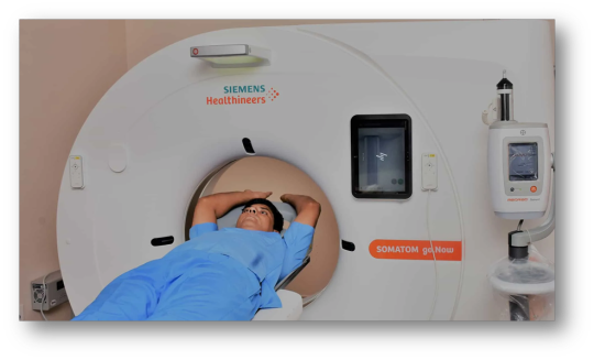
MRI Scan in Ahmedabad
What is MRI ??
MRI, short for Magnetic Resonance Imaging, is a non-invasive medical imaging technique. It is widely used for diagnostic purposes in Ahmedabad and around the world. When seeking the best MRI scan services in Ahmedabad, Imaging World is a prominent name to consider.
Imaging World in Ahmedabad:
Imaging World is recognized for providing high-quality MRI scans in Ahmedabad. Their facility is equipped with state-of-the-art MRI machines that offer exceptional image clarity, aiding in accurate diagnoses. Here are some reasons why Imaging World stands out as one of the best options for MRI scans in Ahmedabad:
Advanced Technology: Imaging World uses cutting-edge MRI technology, ensuring detailed and precise images. This is crucial for diagnosing various medical conditions effectively.
Experienced Radiologists: The center employs experienced radiologists who interpret the MRI images with expertise, contributing to accurate diagnoses.
Patient Comfort: Imaging World prioritizes patient comfort and safety during MRI scans. Their staff is trained to assist patients throughout the process, making it as stress-free as possible.
Wide Range of Services: Imaging World typically offers a broad spectrum of MRI services, including scans of the brain, spine, joints, abdomen, and more.
Timely Reports: Quick and accurate reporting is vital in healthcare. Imaging World strives to provide timely reports to both patients and referring physicians.
Accessibility: Conveniently located in Ahmedabad, Imaging World ensures accessibility for patients from the city and its surrounding areas.
When considering an MRI scan in Ahmedabad, choosing a reputable facility like Imaging World can be a key step in obtaining accurate diagnoses and quality healthcare. It's important to consult with your healthcare provider to determine if an MRI is the appropriate diagnostic tool for your specific medical needs.
We keep the MRI scan cost affordable to meet your financial requirements.
MRI Scan Price Start from : 3600 ₹ onwards after 20% discount on Prior Appointment (Discount code : "PRIOR20")
Book Now
Our Services
MRI Scan in Ahmedabad
CT Scan in Ahmedabad
Sonography in Ahmedabad
Colour Doppler in Ahmedabad
Digital X-Ray in Ahmedabad
2D Echo in Ahmedabad
Contact Information
12A, 14, 15, Ground Floor, Amrapali axiom, near Domino's pizza, besides vakil bridge, Ambli - Bopal Rd, Ambli, Ahmedabad,Gujarat [email protected] +91 70210 76513 02717 488 001/002
#MRI scan price in Ahmedabad#imaging world diagnostic center#CT Guided Biopsy in Ahmedabad#ct scan ambli ahmedabad#CT Scan in ambli ahmedabad#CT Guided Biopsy Bopal#CT Guided Biopsy in Bopal#CT Guided Biopsy ahmedabad#CT Guided Biopsy centre in bopal#USG Guided Biopsy Bopal#USG Guided Biopsy in Bopal#USG Guided Biopsy ahmedabad#USG Guided Biopsy centre in bopal#USG Guided Biopsy in Ahmedabad#mri scan Service bopal#diagnostic centre Bopal#mri scan service Ahmedabad#Colour Doppler in Bopal#2D Echo Bopal#2D Echo in Bopal#mri scan centre in bopal#Digital X-RAY ahmedabad#diagnostic imaging center#CT Scan Bopal#CT Scan in Bopal#Digital X-RAY Bopal#Digital X-RAY in Bopal#Digital X-RAY centre in bopal#CT Scan centre in bopal#2D Echo centre in bopal
0 notes
Text
Ultrascan Diagnostics: Best Diagnostic Centre in Indore for Doppler Scans

Ultrascan Diagnostics is recognized as the best diagnostic centre in Indore, offering high-quality Doppler scans at affordable prices. Our expert radiologists and state-of-the-art equipment ensure accurate results for a variety of Doppler tests, including renal Doppler test in Indore, carotid Doppler test, and penile Doppler test.
Comprehensive Doppler Scan Services
We provide a wide range of Doppler scan services, including:
Renal Doppler Test: Evaluates blood flow to the kidneys and helps diagnose renal artery conditions.
Carotid Doppler Test: Detects blockages and abnormalities in the carotid arteries, reducing stroke risk.
Penile Doppler Test: Assesses blood flow in the penile region to identify erectile dysfunction causes.
Fetal Doppler Scan: Monitors blood circulation in the fetus during pregnancy.
Colour Doppler Ultrasound for Legs: Examines blood flow in the lower limbs to detect deep vein thrombosis (DVT).
Affordable Colour Doppler Ultrasound Price
At Ultrascan Diagnostics, we prioritize affordability without compromising quality. Whether you need a doppler test near me or want to inquire about the color doppler test price, we provide cost-effective solutions. Our pricing is transparent and competitive, ensuring that you receive the best value for your healthcare needs.
Importance of Doppler Scans in Healthcare
Doppler scans are crucial in diagnosing various medical conditions, including:
Pregnancy Monitoring: Color Doppler ultrasound in pregnancy helps monitor fetal health.
Vascular Conditions: Doppler tests detect blood flow issues in arteries and veins.
Cardiovascular Health: Assessing blood circulation to prevent heart-related complications.
FAQs About Doppler Scans
Q1: How much does a color Doppler test cost at Ultrascan Diagnostics? A: The cost of a color Doppler test varies depending on the type of scan required. Contact us for the latest pricing details.
Q2: How should I prepare for a renal Doppler test in Indore? A: It is recommended to fast for 6-8 hours before the test to ensure accurate results.
Q3: Where can I find the best diagnostic centre in Indore for Doppler tests? A: Ultrascan Diagnostics offers comprehensive Doppler scan services with experienced professionals and advanced technology.
Conclusion
Ultrascan Diagnostics is committed to providing the most reliable and affordable renal Doppler test in Indore. Whether you're searching for a doppler scan near me, a fetal doppler scan, or a specialized vascular assessment, our expert team is here to assist you.
If you are searching for a doppler scan near me, look no further than Ultrascan Diagnostics. Contact us today to schedule your appointment and experience top-quality diagnostic care.
Contact Detials [email protected] Mon to Sat : 08:00 AM to 08:00 PM +91 78695 24599, 0731-4100544, 9131492914 Our Location 451-G Greater Brajeshwari,Near Kerala Bakery,Pipliyahana Road, Pipliyahana, indore, Madhya Pradesh 452016
0 notes
Text
Why Lotus Imaging & Diagnostic Centre is the Best Diagnostic Centre Near Me

If you’re looking for reliable health check-ups and tests in Vaishali Nagar, Jaipur, Lotus Imaging & Diagnostic Centre is the place to visit. They are known as the Best Diagnostic Centre in Vaishali Nagar, Jaipur, and here’s why.
Lotus Imaging & Diagnostic Centre uses the latest equipment to get accurate results. Whether you need an MRI, CT scan, or ultrasound, they have the modern machines that give clear images. This helps doctors understand what’s happening in your body so they can provide the right treatment.
But it’s not just about the machines. The staff at Lotus Imaging & Diagnostic Centre are friendly and caring. They make sure you feel comfortable and at ease during your visit. From the moment you walk in, they treat you with kindness and respect.
The centre also makes it easy to get your tests done quickly. They help you set up appointments without any hassle and make sure you get your results in a timely manner. This way, you don’t have to wait long to get the information you need.
In short, if you need a trustworthy place for medical tests, Lotus Imaging & Diagnostic Centre is a great choice. As the Best Diagnostic Centre in Vaishali Nagar, Jaipur, they offer both top-notch technology and excellent care. So, if you want reliable results and a supportive experience, this is the place to go.
This post is tagged in:
Sonography
colour Doppler
Diagnostic centre
Fetal Centre
0 notes
Text
The Comprehensive Ultrasound and Colour Doppler Services at Dokki Scan
In the field of modern diagnostic imaging, the combination of Ultrasound and Colour Doppler technology has become an indispensable tool for healthcare professionals. At Dokki Scan, a renowned medical imaging center in Egypt, patients can access a comprehensive range of Ultrasound and Colour Doppler services that leverage the latest advancements in this transformative technology.
Ultrasound, a non-invasive imaging technique that uses high-frequency sound waves to create detailed images of the body's internal structures, has long been a staple in medical diagnostics. The team at Dokki Scan recognizes the power of this modality and offers a wide array of Ultrasound examinations to cater to the diverse needs of their patients.
From the traditional abdominal and pelvic Ultrasound to the more specialized musculoskeletal, thyroid, and infant brain scans, Dokki Scan's expertise spans a vast spectrum of applications. Patients can also benefit from advanced techniques like 3D and 4D examinations, which provide a more comprehensive and detailed understanding of the body's anatomy and function.
Complementing the Ultrasound services, Dokki Scan's state-of-the-art color Doppler technology takes diagnostic capabilities to new heights. Color Doppler, a specialized Ultrasound technique, allows for the visualization and assessment of blood flow, enabling healthcare professionals to detect and monitor a wide range of vascular conditions.
The comprehensive color Doppler services offered at Dokki Scan include examinations of the carotid and vertebral arteries, the aortoiliac arteries, the upper and lower limb arteries and veins, the renal arteries, and the hepatic vasculature, among others. These advanced imaging tools play a crucial role in the early detection and management of cardiovascular diseases, peripheral vascular disorders, and other related health concerns.
One of the standout features of Dokki Scan's Ultrasound and Colour Doppler services is the integration of cutting-edge biopsy techniques. Patients in need of a more detailed evaluation of focal lesions or suspected abnormalities can undergo Ultrasound-guided biopsies, ensuring a minimally invasive and highly accurate diagnostic approach.
Furthermore, the center's expertise extends to advanced procedures such as Ultrasound-guided aspirations, tappings, and tissue samplings. These specialized services, combined with the advanced imaging capabilities, empower healthcare providers to make well-informed decisions and deliver personalized, evidence-based treatment plans.
Dokki Scan's commitment to providing comprehensive and state-of-the-art Ultrasound and Colour Doppler services is further exemplified by their investment in the latest technological advancements. The center's commitment to innovation ensures that patients have access to the most accurate and reliable diagnostic tools available.
The skilled team of radiologists and technicians at Dokki Scan are trained to interpret the complex data generated by these sophisticated imaging modalities, translating the findings into actionable insights that inform clinical decision-making. This level of expertise, coupled with the center's dedication to patient-centric care, ensures that every individual who walks through their doors receives a tailored and comprehensive diagnostic experience.
In conclusion, Dokki Scan's comprehensive Ultrasound and Colour Doppler services epitomize the future of diagnostic imaging. By combining cutting-edge technology, specialized expertise, and a patient-focused approach, the center has established itself as a beacon of excellence in the field of medical imaging. Whether individuals require routine screening, advanced vascular assessments, or specialized interventions, Dokki Scan is poised to deliver the most accurate and reliable diagnostic solutions, empowering healthcare providers to make informed decisions and improve patient outcomes.
0 notes
Text

Spandan Diagnostic Centre Pvt. Ltd. is one of the top diagnostic centre in Bhubaneswar & one of the leading businesses in the Pathology Labs. It is situated at Plot no.1294 CRPF square, Nayapalli, Bhubaneswar. It is the Largest Health Care Chain Of Eastern And North Eastern Region. Spandan Diagnostic Centre Pvt. Ltd. is built on four major founding factors, which plays a key role in delivering the best of possible care to each and every patient. Each factor takes care of individual needs of patients, thereby enabling customised care for everyone according to their needs and health conditions. At Spandan they have 3.0 T MRI Scan, Cardiac CT Scan, BMD/DXA Scan, USG/Colour Doppler, Full Room DR (X-Ray) & Advanced Pathology.
For more info visit: https://spandandiagnosticcentre.com/
0 notes
Text
Best Ultrasound Centre in Panchkula | Kior Healthcare
Experience top-notch medical imaging services at Kior Healthcare, your trusted destination for mammography in Panchkula, the best CT scan center in Panchkula, ultrasound in Panchkula, and Colour Doppler tests in Mohali. Our state-of-the-art facilities, skilled professionals, and commitment to accuracy ensure precise diagnostics for your health needs. Elevate your healthcare experience with advanced technology and personalized care. Choose Kior Healthcare for excellence in mammography, CT scans, ultrasound, and Colour Doppler tests, setting a benchmark for comprehensive and reliable diagnostic services in the Panchkula and Mohali regions. Your well-being is our priority at Kior Healthcare.

0 notes
Text
Lupine Publishers | Radiology; USG and Colour Doppler of Post Renal Transplant Complications

Abstract
Kidney transplant is the treatment of choice for patients with end-stage renal disease. Kidney transplant offers better quality of life. It is more cost effective than hemodialysis. Advances in surgical technique, along with improvement in organ preservation and immunosuppression have improved patient outcomes. Post-operative complications, however, can limit this success. Ultrasound and Doppler study is the primary imaging modality for evaluation of renal transplant, providing real –time information about complication in graft. A multimodality approach including CT scan, MRI or conventional angiography may be necessary in cases when sonography and Doppler are inconclusive to diagnose the etiologies of these complications. Radiologists play an integral role within the multidisciplinary team in care of transplant patient at every stage of the transplant process. Therefore, the radiologist should always be aware when evaluating the failing renal graft, whether the cause is renal or extrinsic. In this pictorial essay we tried to gather the most common complication of transplant kidney in different cases that occurred in our hospital, with an emphasis on Ultrasound and Doppler study.
Keywords: USG; Colour Doppler; Post renal transplantation; Complications
Introduction
The preferred modality for renal replacement is renal transplantation, and its superiority in prolonging the longevity of patients with end-stage renal disease is well established [1]. Kidney transplantation is typically classified as deceased-donor (formerly known as cadaveric) or living-donor transplantation depending on the source of the donor organ. Due to improvement in transplantation technology, advancement in immunosuppressant and graft preservation techniques the 1-year survival rates for grafts, are reported to be 80% for mismatched cadaveric renal grafts; 90% for nonidentical living related grafts; 95% for human lymphocyte antigen-identical grafts [2]. Radiologists play a major role at every stage of the transplant process with transplantation team. Ultrasonography with colour Doppler is the principal modality used for evaluation of renal allograft. USG is a relatively cheap, noninvasive, and non-nephrotoxic modality. It is applied for diagnostic and monitoring purposes in the post-transplant period. This pictorial essay describes USG and Doppler imaging appearances of the major complications that may occur in renal transplantation. All our images have been obtained from a single our center which is major transplantation center in India. All post renal transplant patients undergo a USG and comprehensive Doppler evaluation on post-operative day one. The sonographic examination algorithm includes gray-scale evaluation of the graft and spectral Doppler. Further imaging is based on clinical follow-up including daily monitoring of laboratory values. If clinical parameters are abnormal, repeat sonography is performed and depending on the results, CT, MRI, or angiography may be requested.
Surgical Technique
Surgical technique and location of placement of renal allograft depends on the variation in arterial and venous anatomy of donor. The transplanted kidney is usually placed extraperitoneally in the patient’s right iliac fossa (less commonly in left iliac fossa), with end-to-side or end to end anastomosis to the external iliac vasculature. The currently preferred method for restoring urinary drainage is ureteroneocystostomy, a procedure by which the ureter is implanted directly into the dome of the bladder (Figure 1).
Urologic Complications
The prevalence of urologic complications varied from 10% to 25% with a mortality rate ranging from 20% to 30%. Incidence rate is decreased range between 3% and 9% in the current era because of advancement in surgical techniques and frequent use of ureteral stents [3,4].
Urinary Obstruction
a) Incidence: 2%-5% of kidney transplant recipients.
b) Site of obstruction: Approx. 90% of stenoses occur in the distal third of the ureter due to its poor vascular supply.
c) Imaging appearance: US can easily confirm the diagnosis of hydronephrosis and dilated renal pelvis and thus determine the level of ureteral obstruction (Figure 2). When contents of pelvic calyceal system are echogenic and weakly shadowing, fungus balls should be considered, whereas low-level echoes suggest pyonephrosis (Figure 3).
Urine Leaks and Urinomas
a) Incidence: up to 6 % of renal transplant recipients [5]
b) Location: extraperitoneal or intraperitoneal, if intraperitoneal may present with ascites.
c) Imaging appearance: USG findings are nonspecific, well defined anechoic fluid collection with septa or without septation, adjacent to the lower pole of the transplant in most of the cases (Figure 4).
Drainage of fluid under ultrasound guidance and testing the fluid for creatinine helps to differentiate it from seromas or lymphoceles. High concentration of creatinine will be found in case of urinoma if we compare with serous fluid.
Calculous Disease
a) Incidence: 1% to 2 % of post-transplant cases develops clinically relevant stones as compared to general population [6]. As the kidney is denervated patient will not suffer typical renal colic.
b) Imaging appearance: Calculus appears as strongly reactive focus of variable size producing acoustic shadowing on USG and twinkling artifact on colour Doppler (Figure 5). Other rare urologic complications are ureteric necrosis and vesico-ureteric reflux.
Peritransplant Fluid Collections
Fluid collection in peritransplant region has been reported in up to 50 % of renal transplantation. They are urinomas, hematomas, lymphocele and abscess, the clinical relevance of these collection is largely determined by their size, location and possible growth. In immediate postoperative period, small hematomas or seroma are almost expected. Their size should be documented at baseline examination. Rarely do they lead to graft dysfunction or obstruction of collecting system. Urinomas and hematomas are most likely to develop immediately after transplantation, whereas lymphoceles generally develop after 4 to 8 weeks. The ultrasonography characteristics of peritransplant fluid collections, however, are entirely nonspecific, correlation with clinical findings helps to restrict differential diagnosis. Ultimately, diagnosis may be made only with percutaneous aspiration and then biochemical analysis. Differentiation between Peritransplant and subcapsular collection is important. A subcapsular collection likely to cause mass effect on parenchyma, usually cresenteric and show extension along the contour of kidney deep to the renal capsule
Hematoma
a. Incidence: Varies from 4 to 8 % [7]
b. Imaging appearance: Hematomas have a complex appearance. Hematomas appears echogenic in acute case and progressively become less echogenic with time (Figure 6). Chronic hematomas even appear anechoic, more closely resembling fluid and septation may develop later on.
Lymphocele
a. Incidence: Affecting up to 20% of the patients [8] It occurs after surgery owing to the surgical disruption of the normal lymphatic channels along the iliac vessels or around the hilum of the graft.
b. Imaging appearance: on Ultrasound it appears as anechoic bur may contain septation. They may become infected and gave more complex appearance (Figure 7).
Peritransplant abscesses
a) Imaging appearance: USG cannot always differentiate an abscess from other collection. Collection may show low level echoes and thick irregular wall. If gas is seen, an abscess is probable. In any pyrexia patient, any perinephric collection should be considered infected until proven otherwise through the appropriate imaging and guided diagnostic aspiration.
Parenchymal Complication / Graft Dysfunction
Diseases of the renal parenchyma are usually diffuse and often leads graft dysfunction. Differential diagnosis is difficult by imaging alone. USG is not sensitive or specific for evaluation; differential will be relying on biopsy. USG still has a central role in qualitative assessment of graft perfusion and to guide the biopsy.
Acute tubular necrosis (ATN)
Acute tubular necrosis is due to reversible ischemic damage to the renal tubular cells prior to engrafting and reperfusion injury.
i. Incidence: affects 20–60 % of cadaveric renal grafts.
ii. Time of onset: in the first 48 hours after transplantation.
iii. Imaging appearance: No specific imaging pattern for the diagnosis of ATN. The kidney may appear normal, in severe cases it looks enlarged, edematous and echo poor with loss of corticomedullary differentiation (Figure 8) and shows elevated RI (above 0.8).
Rejection
Rejection can be classified according to the period of appearance as hyper acute (occurring within minutes), acute (occurring within days to weeks), late acute (occurring after 3 months), or chronic (occurring months to years after transplantation). Classification of renal allograft rejection by Banff classification of allograft pathology is routinely followed nowadays. It is based on a combination of histopathologic features coupled with molecular, serologic, and clinical parameters.
Acute Rejection (AR)
i. Incidence: up to 40% of patients in the early transplant period [9].
ii. Imaging appearance: Graft enlargement due to edema, Decreased cortical echogenicity, swelling of the medullary pyramids, echogenic sinus fat, edematous wall of pelvic calyceal system, focal hypoechoic areas in parenchyma which may favors infarct and collection in perigraft region due to necrosis or hemorrhage. PI and RI elevated in both ATN as well as in AR, but AR has high values of it. In severe cases, Power Doppler shows reduced, absent or reversed diastolic flow with elevation of the RI (Figure 9).
Chronic Rejection
Chronic rejection occurs in case of insufficient immunosuppression given to recipient to control the residual antigraft lymphocytes and antibodies.
i. Imaging appearance: US appearance is not typical, ranging from normal to hyper echogenic, along with cortical thinning, reduced number of intrarenal vessels, and mild hydronephrosis (Figure 10). RI measurements are not reliable for this diagnosis. The diagnosis is made histologically.
Drug Nephrotoxicity
Calcineurin inhibitors are key immunosuppression agents administered to avoid acute rejection, but they are nephrotoxic.
A. Imaging appearance: USG may show completely normal results or nonspecific findings such as graft swelling, increased or decreased renal echogenicity and loss of cortico-medullary differentiation. Doppler study may show a RI increase of 0.80. Findings of USG and Doppler study should be correlated with the serum drug levels. USG and Doppler findings of ATN and AR is almost similar, but both can be differentiated by time course of the findings. Clinical & biochemical correlation and serial measurements of RI and Pulsatile index (PI) would be further helpful to monitor the patient.
Infections and abscesses
Incidence and time of onset: More than 80% of renal transplant recipients have at least 1 episode of infection during the first year of post transplantation.
I. Imaging appearance: USG appearance is quite variable. Focal pyelonephritis appear as a focal hyperechoic or hypoechoic area, but this finding is nonspecific because it can represent infarction or rejection also (Figure 11). Abscess has varied appearance on USG like- heterogeneous, hypoechoic or cystic. Urothelial thickening may be seen. In febrile post renal transplant patient low level echoes in dilated collecting system may favors pyonephrosis. Fungus ball appears as focal rounded, weakly shadowing or echogenic structure in dilated pelvic calyceal system. In emphysematous pyelonephritis, gas in the parenchyma of the renal graft produces an echogenic line with distal reverberation artifacts. Papillary necrosis has no typical sonographic findings, and it subsequently leads to ureteric obstruction.
End stage disease
Nonfunctional renal grafts are left in place in abdominal cavity. Gradually graft becomes small and can have fatty replacement, hydronephrosis, infarcts, hemorrhage or calcification.
Vascular Complications
Vascular complications in post renal transplantation have significant negative influence of graft survival. They are infrequent, occur in approximately 1%���2%; [10] but can cause sudden loss of renal allograft. Selective catheter angiography is the gold standard for diagnosis; however, it is invasive and may cause various complications. Hence it is not used as a screening tool but reserved for patients with inconclusive results on the noninvasive screening tests. Noninvasive imaging like ultrasound, Doppler, scintigraphy, CT and MR angiography plays major role to evaluate them.
Renal artery thrombosis (RAT)
Incidence: Ranges from 0.5% to 3.5 % [11]
Imaging appearance: No evidence of any arterial or venous flow noted on color, spectral and power Doppler study (Figure 12). Doppler sonography had 100% sensitivity and specificity for diagnosis and hardly any other imaging study is required for diagnosis [12].
Focal Renal Infarction
Imaging appearance: A segmental infarct appears as a poorly marginated wedge shaped hypoechoic mass or a hypoechoic mass with a well-defined echogenic wall without colour flow (Figure 13). If the infarction is global, the kidney will appear hypoechoic and be diffusely enlarged.
Renal Artery Stenosis (RAS)
Incidence: It has wide range varying from 1% to 23 % depending on the definition and diagnostic techniques used.
Site: usually at anastomotic site
Imaging appearance: On gray scale USG, there is lack of normal post-transplant hypertrophy. On color Doppler study appearance of focal color aliasing noted at stenotic segments. On spectral Doppler study, peak systolic velocity in main renal artery >300 cm/sec and Ratio of PSV in transplanted main renal artery and external iliac artery greater than or equal to 1.8 are highly suggestive of significant stenosis. Indirect criteria are low resistive index <0.56, Acceleration time >0.07 sec, Acceleration index <3 meter/ sec and Intrarenal tardus–parvus waveform (Figure 14).
Limitation: Results are strongly depends on the operator’s individual experience and skill.
Rate of restenosis is less than 10 %. Doppler ultrasonography is the procedure of choice to evaluate graft perfusion before and after revascularization The term pseudo transplant renal artery stenosis (TRAS) refers to thrombosis or stenosis of iliac artery or aorta proximal to transplant renal artery.
Renal vein thrombosis: (RVT)
Incidence: Ranges from 0.9% to 4.5 % [13]
Imaging Findings: Graft appears swollen and hypoechoic.
Doppler shows absent venous flow. Renal arterial Doppler spectrum shows absent or reversal of diastolic flow due to increased resistance (Figure 15). Reversal flow in renal artery is nonspecific as it also seen in severe rejection and in acute tubular necrosis, its combination with absent venous flow is the diagnosis of renal vein thrombosis. Partial thrombosis also can occur near anastomosis or within the transplanted kidney (Figure 16).
Extra parenchymal pseudo aneurysm
Incidence: Anastomotic pseudoaneurysm is a rare complication of renal transplantation occurring in 0.3%. [14]
Imaging findings : On gray scale ultrasound it appears as cystic lesion which shows color flow and to and fro spectral pattern on doppler study(Figure 17). Endovascular treatment with covered stent placement to exclude pseudoaneurysm can also be evaluated with USG and colour doppler (Figure 18).
Intra-parenchymal arteriovenous fistula and pseudoaneurysm
AVF occurs when both artery and vein are simultaneously lacerated, while pseudoaneurysm results when only artery is lacerated.
Incidence: 1-18% of the biopsies [15]
Time of onset: occur at time of biopsy. They depend on many factors – ultrasound guidance, needle caliber and imaging follow up.
Imaging appearance: colour Doppler study shows AVF as focal areas of disorganized flow adjoining the normal vasculature. Spectral waveforms show increased arterial and venous flow with high velocity and low resistance (Figure 19). Pseudoaneurysm appears as simple or complex cyst on B mode ultrasound and intracystic flow on colour Doppler mode (Figure 20).
Neoplasms after renal transplantation
Post renal transplantation patients are at higher risk of development of neoplasms. Urologic tumour are 4 to 5 times more common in post renal transplantation recipients than normal population with significant exposure to cyclophosphamide immunosuppressant agent.
Renal cell carcinoma
Etiology: by means of transplanted organ or de novo development by immunocompromised status, patients on hemodialysis in case of chronic renal failure develop acquire renal cystic disease
Prevalence: 90 % occurring in native kidney and 10 % in transplanted kidney [16]
Imaging appearance: lesion appear heterogeneous with vascularity, similar picture as seen in native kidney [17] (Figure 21).
Lymphomas
Incidence: 1 % of renal allograft recipients [18]
Time of onset: Post transplantation lymphoproliferative disorder is diagnosed at a median of 80 months after transplantation.
Imaging appearance: Lymphadenopathy at various sites but can also affect any solid organ or hollow viscera or transplant graft parenchyma itself. It appears as low or mixed reactive masses and tends to have a predilection for the renal hilum.
Recurrent Native disease
It depends on the primary disease before transplantation.
Imaging appearance: Imaging has no specific pattern in these situations apart from excluding the treatable cause of reduced renal function.
Abdominopelvic Complications
Renal graft is placed in extraperitoneal space via a peritoneal window in laparoscopic and robot assisted surgical techniques. So these cases are prone to same complications experienced by other surgical cases in whom peritoneum is exposed.
Renal Allograft Compartment Syndrome (RACS)
It is a rare syndrome, and it is under recognized cause of early transplant dysfunction or even loss. It may occur as a result of intracompartment hypertension and ensuring ischemia of renal graft [19]. Imaging appearance: absent or diffuse diminished cortical flow in transplant kidney at colour Doppler.
Fascial dehiscence and bowel or allograft evisceration
They tend to occur in perioperative period. Herniation of bowel through a transplant peritoneal defect may lead to compromise of intestine or transplant itself.
Limitations
The USG examination is examiner dependent and limited accessibility in obese patents impairs the evaluation and often leads misinterpretation. The RI index is also unspecific and influenced by many factors like site at which the RI is measures, increased intraabdominal pressure during forced inspiration and the pulse rate.
Summary
Kidney transplant is the treatment of choice for patients with end-stage renal disease. Improvements in surgical techniques and advanced immunosuppressive drugs have resulted in remarkable survival of patients and renal grafts. Still complications occur in both the early postoperative period and later. Kidney transplants follow up is common in radiology and sonography practice.
Ultrasonography and Doppler examination can accurately depict and characterize many of the potential complications of renal transplantation. It facilitates prompt and accurate diagnosis and thus guiding treatment.
For more Lupine Journals please click here: https://lupinepublishers.com/index.php
For more Journal of Urology & Nephrology Studies articles please click here: https://lupinepublishers.com/urology-nephrology-journal/index.php
#lupine publishers#open access journals#urology#nephrology#submission#journal of urology & nephrology studies#juns#lupine journals#nephritis#articles
0 notes
Text
#diagnostic centre in chandigarh#mri scan in chandigarh#ultrasound services in chandigarh#3d and4d ultrasound centre in chandigarh#colour doppler ultrasound services in chandigarh#digital x-ray services in chandigarh
0 notes
Text
CT Scan Centre in Ahmedabad - Imaging World
Computed Tomography, also known as CT or CAT Scan, it is a diagnostic procedure used to produce detailed X-ray images of the body to detect and reveal the details of internal organs and any health issues that cannot be seen by conventional X-rays. It can produce 2D cross-sectional images of the body’s tissues, bones, and blood vessels.
Gujarat's vibrant metropolis, Ahmedabad, is well-known for its lively marketplaces, extensive history of culture, and first-rate medical facilities. CT Scan Price in Ahmedabad is home to a number of modern facilities offering CT scans and other diagnostic imaging services. These facilities are run by highly qualified specialists and include state-of-the-art equipment. We'll examine some of the top CT scan facilities in Ahmedabad in more detail in this guide, highlighting their offerings, advanced equipment, and patient-centred culture.
CT scan Procedure:
After escorting you to the CT scan room by a technician, you will be asked to lie down on a padded table that can slide in and out of the scanner machine and remain still.
When the pictures are picked up by the X-ray detectors placed in the device and relayed to a computer for processing, you could hear a clicking sound.
After the CT scan Centre in Ahmedabad , you can usually resume your normal activities unless instructed otherwise by your healthcare provider. The images will be processed by a radiologist, who will interpret the results and prepare a report for your referring physician. CT Scan Centre in Bopal, CT scan changodar, CT scan thaltej, CT scan in thaltej.
The CT scanner is a large, doughnut-shaped machine that houses an X-ray tube and detectors. It rotates around your body while capturing multiple X-ray images from different angles. These images are then processed by a computer to create cross-sectional slices, or "slices," of the body.
Our Services: - CT scan Centre
MRI Scan in Ahmedabad
CT scan in Ahmedabad
Sonography in Ahmedabad
Colour Doppler in Ahmedabad
Digital X-Ray in Ahmedabad
2D Echo in Ahmedabad
Contact us: - +91 9909644344, [email protected]
Address: - 12A, 14, 15, Ground Floor, Amrapali axiom, near Domino's pizza, besides vakil bridge, Ambli - Bopal Rd, Ambli, Ahmedabad, Gujarat
Visit for More Information: - https://www.imagingworld.in/
#CT Scan Centre in Ahmedabad#CT Scan Price in Ahmedabad#CT Scan in Bopal#CT Scan Centre in Bopal#CT Scan changodar#CT Scan Sarkhej#CT Scan in Sarkhej#CT Scan thaltej#CT Scan in thaltej
0 notes
Text
Ultrascan Diagnostics: Affordable Color Doppler Ultrasound in Pregnancy in Indore

Looking for a reliable "doppler test near me"? Ultrascan Diagnostics offers high-quality "colour doppler ultrasound in pregnancy" services at an affordable "colour doppler ultrasound price". Our advanced imaging technology ensures accurate results, whether you need a "renal doppler test," "carotid doppler test," or a "fetal doppler scan." Contact us for the best "doppler scan price" in Indore.
Contact Detials [email protected] Mon to Sat : 08:00 AM to 08:00 PM +91 78695 24599, 0731-4100544, 9131492914 Our Location 451-G Greater Brajeshwari,Near Kerala Bakery,Pipliyahana Road, Pipliyahana, indore, Madhya Pradesh 452016
0 notes