#Histomorphometry
Explore tagged Tumblr posts
Visit Tumblr Blog
Explore Tumblr blogs with no restrictions, modern design and the best experience.
Last Seen Tumblr Blogs


Fun Fact
Tumblr was the first site to host the blog for President Barack Obama in 2011.
Link
1 note
·
View note
Photo

Simplify Medical coding Institute. Online Courses used. Standard and Advanced Medical coding. CPC Exam training. Whatsapp: +91936095154 4. Pathology CPT codes, 19459006 Surgical pathology CPT codes, Cpt code for Decalcification treatment, 19459006 Cpt code for Pathology assessment throughout surgical treatment, Cpt code for Electron microscopy, Cpt code for Morphometric analysis, 19459006 Cpt code for Histomorphometry,…
0 notes
Link
Abstract
The decline of circulating testosterone levels in aging men is associated with adverse health effects. During studies of probiotic bacteria and obesity, we discovered that male mice routinely consuming purified lactic acid bacteria originally isolated from human milk had larger testicles and increased serum testosterone levels compared to their age-matched controls. Further investigation using microscopy-assisted histomorphometry of testicular tissue showed that mice consuming Lactobacillus reuteri in their drinking water had significantly increased seminiferous tubule cross-sectional profiles and increased spermatogenesis and Leydig cell numbers per testis when compared with matched diet counterparts This showed that criteria of gonadal aging were reduced after routinely consuming a purified microbe such as L. reuteri. We tested whether these features typical of sustained reproductive fitness may be due to anti-inflammatory properties of L. reuteri, and found that testicular mass and other indicators typical of old age were similarly restored to youthful levels using systemic administration of antibodies blocking pro-inflammatory cytokine interleukin-17A. This indicated that uncontrolled host inflammatory responses contributed to the testicular atrophy phenotype in aged mice. Reduced circulating testosterone levels have been implicated in many adverse effects; dietary L. reuteri or other probiotic supplementation may provide a viable natural approach to prevention of male hypogonadism, absent the controversy and side-effects of traditional therapies, and yield practical options for management of disorders typically associated with normal aging. These novel findings suggest a potential high impact for microbe therapy in public health by imparting hormonal and gonad features of reproductive fitness typical of much younger healthy individuals.
2 notes
·
View notes
Text
Metformin Reduces the Extent of Varicocele-Induced Damage in Testicular Tissue
Authored by: Erkan Erdem*
Introduction
Varicocele is an abnormal vascular dilatation of pampiniform plexus, commonly developing at puberty. Although underlying mechanisms remain poorly understood genetic background, anatomical aberrations, incompetence of venous valves, difference between the drainage of left and right testicular veins were suggested in the etiology [1]. As left spermatic vein being longer than the right vein, it is more commonly incurred to increased hydrostatic pressure and dilatation. Compression of the left renal vein between the aorta and the superior mesenteric artery may also contribute to the disturbed intravenous pressure [2].
The prevalence of varicocele varies between 15-20 % in general population and 30-40% in infertile men, and 11-19% of adolescents [3-6]. It was reported that varicocele is a progressive disease and early diagnosis and treatment in youth may enhance fertility potential [7]. Several contributing factors in the pathophysiology of varicocele have been proposed such as higher temperature of testis, the disorder of neuroendocrine system, autoimmunity, accumulation of renal and adrenal metabolites, genetic and epigenetic factors, hypoxia and oxidative stress [8-10].
Varicocele represents a chronic process within the testicle, which is linked to increased reactive oxygen species (ROS) beyond physiologic limits and, subsequently, disrupting sperm membrane fluidity, causing DNA damage and necrosis [11]. Moreover, superoxide dismutase 1, glutathione S-transferase M1 and T1 which are counteracting free superoxide radicals in cells have been reported to be decreased in men with varicocele, that may be important on disturbed sperm parameters [12]. Apoptosis of germ cells was also demonstrated in the pathogenesis of varicocele-related infertility [13]. Clinical findings suggest that surgical repair of varicocele may decrease seminal oxidative stress levels and sperm DNA fragmentation and, thus, may improve sperm quality [14]. Therefore, surgical intervention seems to be a reliable option in the treatment of varicocele-related male infertility, although some controversial reports exist.
Additionally, anti-oxidant medications such as kallikrein, L-carnitine with L-acetyl carnitine, pentoxifylline, coenzyme Q10 have been used to improve the milieu in the testis in men with varicocele [15]. Metformin is a major therapeutic agent in the treatment of type 2 diabetes mellitus as an insulin sensitizer, which decreases hepatic glucose output and increases peripheral glucose uptake. Although its action was not fully elucidated, metformin attenuated intracellular reactive oxygen species and apoptosis in aortic endothelial cells, myocardium, renal tubular cells and testicular cells [16-20].
Aim
Potential effects of metformin on varicocele-induced testicular damage have not been studied in neither humans nor in animal models. Thus, we investigated the impact of metformin on spermatogenesis, testicular integrity, and apoptotic activity in the testis of adolescent rats with experimentally-induced varicocele.
Materials and Methods
Thirty-six male adolescent Wistar rats (6-week-old) were randomly and equally divided into six experimental groups. Surgical procedures were carried out under anesthesia with intraperitoneal injection of ketamine (50 mg/kg). The experimental groups were as follows:
• (C) Control group; no surgical procedure was performed, and testis was examined after removal.
• (S) Sham group, a midline incision was performed, and testis was examined 8 weeks later.
• (V) Varicocele - only group: Experimental varicocele was induced by partial ligation of left renal vein with
Silk suture at the area medial to the insertion of the adrenal and spermatic vein into renal vein as described previously [21].
• (V+M) Varicocele + metformin group: All rats were treated with metformin (300 mg/kg per day by oral gavages) for 8 weeks following induced varicocele.
• (V/E) Varicocele + varicocelectomy group: Varicocelectomy was performed 4 weeks and the examination of the testis 8 weeks after the induction of varicocele. No medication was used.
• (V/E+M) Varicocele + varicocelectomy + metformin group: Varicocelectomy was performed 4 weeks after the induced varicocele. Metformin treatment (300 mg/kg per day by oral gavages) was initiated after the induction of varicocele and continued for 8 weeks. Left testes were examined 8 weeks after the induction of varicocele in all varicocele - induced groups. As maximum apoptotic activity initiates approximately 28 days after the induction of varicocele the procedure of varicocelectomy was performed 4 weeks after the formation of varicocele [22].
Histologic preparation and evaluation
The testicular tissue was fixed in Bouin’s solution (75% picric acid, 5% glacial acetic acid, and 25% formaldehyde) and embedded in paraffin blocks. Sections (5 μm) were formed, deparaffinized, and stained with hematoxylin and eosin. Spermatogenesis was examined in each group using Johnsen’s score (a score of 1-10 was assigned to each tubule regarding epithelial maturation) as described previously [23]. Sections were examined in a random order under a standard light microscope with 10x and 40x magnification by a blinded histologist; unaware of which group each rat belonged to. Histological grading was done by examining approximately 80 randomly selected seminiferous tubules per rat. Thus, a total of approximately 480 seminiferous tubules were scored for each group.
Histomorphometry analysis
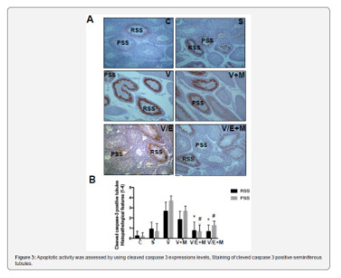
A total of 103 randomly selected seminiferous tubules stained with hematoxylin-eosin were analyzed in each group. The presence of round spermatid stage (RSS) and primary spermatocyte stages (PSS) were assessed as described previously and compared among the groups [24].
Immunohistochemical staining for cleaved caspase-3 and ImageJ analysis
Cleaved caspase-3 was used for immunohistochemical staining. Testicular tissue samples were immediately fixed in 10% neutral-buffered formalin, embedded in paraffin, and sectioned (5 μm). Sections were deparaffinized and blocked for endogenous peroxidase activity with methanol containing 3% H2O2 for 10 m. Ultra V Block (Lab vision, Freemont, CA) for 7 m at room temperature. Cleaved Caspase-3 (#9664, Cell Signaling, U.S.) was applied at a dilution of 1: 500 and incubated overnight at +4 °C in a humidified chamber for nonspecific binding. The sections were washed in phosphate-buffered saline (PBS) and incubated with biotinylated horse anti-rabbit IgG (3 mg/mL; Vector, Burlingame, CA) at a 1: 500 dilution for 1 h at room temperature.
Antibodies were detected using a VECTASTAIN avidinbiotin complex (Vector PK 4000) for 30 m at room temperature. Antibody complexes were visualized after incubation with 3,3’-diaminobenzidine tetrahydrochloride (DAB, Bio-Genex, San Ramon, CA.) and were mounted under glass coverslips in Entellan (Merck) and then evaluated under a light microscope. Immunohistochemical staining for cleaved caspase-3 was analyzed by counting 100 seminiferous tubule cross-sections in each group and expressed as the apoptotic index. In each photomicrograph, the following parameter was measured with ImageJ software: expression levels of cleaved-caspase-3 in both groups at round spermatid stage (RSS) of testes. Each of this parameter was measured 3 times for each image and the average of the 9 measurements of each sample was used for the statistical analysis. Histopathological features examined in rats with normal testis and with sham, varicocele, varicocele+ metformin in a subjective scoring (0 - not present; 1 - low grade; 2 - moderate grade; 3 - high grade; 4 - very high grade).
Statistical analysis
Histopathological findings (Johnsen’s score) were assessed by nonparametric Kruskal-Wallis test, and the mean Johnsen’s score was used in the comparison of the groups. Multiple comparisons were made using Tukey’s procedure. p<0.05 was considered statistically significant. Analysis of variance was used for statistical analysis of the apoptotic index among the groups.
Results
Assessment of spermatogenesis
Johnsen’s score was significantly lower in V group (4.14±1.25) compared to C group (9.1±0.3) or S group (9.0 ± 0.2) groups (p<0.05). V+M group had significantly higher score (6.9±0.6) than V group (p<0.05). V/E group and V/E+M group had similar Johnsen scores (8.9 ± 1.02 and 9.2 ± 0.6). These findings suggest that the administration of metformin resulted in 40.6% of improvement in spermatogenesis in rats with varicocele. However, this favorable effect was not observed when metformin was used along with varicocelectomy.
Histological and morphological changes in seminiferous tubules
Histological and morphological changes in the testes of rats were compared via hematoxylin and eosin staining and degenerated tubules (DT) were only detected in V and V+ M groups, not in C, S, V/E and V/E+M groups (Figure 1). Visual assessment of the disorganized seminiferous tubules further supported these findings as seen in Figure 2. Seminiferous tubule degeneration scores were used for quantification of data (Figure 2b). V group had significantly higher scores of RSS and PSS compared to C and S group (2.6±0.8 and 3.7±0.4; 0.2±0.4 and 0.2±0.4; 0.9±0.6 and 0.6±0.7, respectively) (p<0.05). V/E group had significantly lower RSS (0.7±0.8) and PSS (0.8±0.7) scores than V group (p<0.05). V+M group had significantly lower RSS and PSS scores (1.8±0.7 and 2.6±0.7, p<0.05) in comparison to V group, implicating beneficial effects of metformin in rats with varicocele. When compared to V/E group, V/E+M group did not exhibit any difference in RSS (0.6±0.6) and PSS (1.4±0.5) scores, suggesting the absence of additive positive effect of metformin in varicocelectomies rats.
Apoptotic activity
Apoptotic activity was assessed by using cleaved caspase 3 expressions levels, staining of cleved caspase 3 positive seminiferous tubules were shown in Figure 3a. Cleaved caspase 3 expressions were significantly higher in V group (3.5 ± 0.5) compared to C (0) and S (0.2 ± 0.4) groups. V+M group had significantly lower cleaved caspase 3 level (3.0 ± 0.7) than V group. V/E group had lower cleaved caspase-3 expression levels (1.0 ± 0.7) compared to V group. Treatment of varicoceleectomy rats with metformin (V/E+M) did not further reduce apoptotic activity in the seminiferous tubules (1.75 ± 0.43) when compared to the varicocelectomy group (V/E) (Figure 3b).
Discussion and Conclusion
The present study demonstrates that metformin can reduce the extent of testicular damage in rats with varicocele, although having no effect in rats following varicocelectomy Spermatogenesis, seminiferous tubule integrity and the degree of apoptosis were improved using metformin in the presence of varicocele although it was not as remarkable as what was obtained through varicocelectomy. A review of the literature revealed that the impact of metformin on varicocele was not investigated in humans or animal models until now.
Although it is a commonly identified abnormality not all men with varicocele present with infertility. Some intrinsic factors may render some men to become susceptible to varicocele, thus, the best candidates who benefit from varicocelectomy yet to be clarified. Since oxidative stress was shown to be important in the pathophysiology of varicocele some agents have been used to improve the milieu in the testis [1]. A number of anti-oxidant medications have been studied to relieve detrimental effects of varicocele in the testis [25]. These agents have been used either alone or as an adjuvant therapy with surgery. However, surgery remains the treatment of choice and there exists insufficient data to recommend medical therapy in men with varicocele. Barekat et al. [26] reported that administration of an antioxidant agent N-acetyl cysytein as an adjunct therapy improved semen quality following varicocelectomy [26]. Tek et al. [21] demonstrated that vascular endothelial growth factor decreased apoptosis in varicoceleinduced rats as evidenced by diminished caspase-3 positive cells [21]. Both studies showed the benefit of these as adjunct therapy following varicocelectomy. However, in the present study metformin did not enhance the effect of varicocelectomy.
Minutoli et al. [13] demonstrated that neuronal apoptosis inhibin factor and surviving expressions were significantly reduced following varicocele induction and polydeoxyribonucleotide, an agonist of adenosine A2A receptor, administration restored testicular function [13]. Several other studies detected increased germ cell apoptosis in rats with varicocele [21,22,27]. However, in another study, apoptosis was found to be decreased in germ cells in the testes of infertile men with varicocele as compared with normal men [28]. It was speculated that the fixation of testis in formaldehyde might have played a role in the different result. In the present study, cleaved caspase 3 expression was used to assess apoptotic activity and it was found that metformin reduced apoptotic activity in rats with varicocele, whereas no additional effect was observed when metformin was administered after varicocelectomy.
Metformin is commonly used in type 2 diabetes mellitus and polycystic ovarian disease as an insulin sensitizer [29]. Also, it is present in various tissues including myocardium, liver, pancreas, thyroid, adipose tissue, hypothalamus, pituitary, and male and female gonads [19,30,31]. It has been reported that metformin is mainly transported into cells by organic cation transporters as passive diffusion is limited [32]. Although the mechanism of action is not yet fully elucidated recent studies suggested that metformin acts through AMP-activated protein kinase (AMPK) pathway, inhibits the activity of the respiratory electron transport chain in mitochondria, induces epigenetic modifications which in part may explain long term effects and decreases oxidative stress and apoptotic activity [16,19, 33-35].
Male reproductive system utilizes all these metabolic pathways and is prone to be affected by metformin administration [20,36,37]. Metformin was found to stimulate lactate production which is important in the development of germ cells and show an anti-apoptotic effect in rat Sertoli cells [38]. It was also reported that metformin reduced the apoptotic cells and caspase-3 level in rat testis [20]. The findings of the present study are consistent with previous studies that metformin reduced apoptosis in testis with varicocele. Yan et al. [37] reported that metformin improved the semen parameters related to its effects on weight loss, increased testicular weight and reduced testicular cell apoptosis [37]. On the other hand, Tartarin et al. [36] reported metformin at concentration 10 times higher than therapeutic levels decreased testosterone secretion and the number of Sertoli cells in rats when it was administered during pregnancy [36]. Faure et al. [39] reduction in testicular weight and testosterone level were observed in 6-week-old chickens treated with metformin for 3 weeks [39].
Several groups demonstrated that post-operative administration of metformin can exert protective effects in male reproductive function in rat models [40]. Bosman et al. [41] demonstrated that infertile hyperinsulinaemic men could benefit from metformin treatment in combination with an enriched antioxidant diet [41]. Besides, metformin was reported to act as a protective compound when used in the media for cryopreservation of spermatozoa [30]. In conclusion, metformin reduces detrimental effects of varicocele, although no additional benefit is expected following varicocelectomy. Further studies are required to apply metformin for this indication in humans.
#open access journals#peer review journals#Juniper Publishers PubMed Indexed Articles#reproductive medicine#GJORM in juniper publishers
1 note
·
View note
Text
Reconstruction of Large Abdominal Wall Defects Using Neurotized Vascular Composite Allografts
Karim Sarhane MD
Background: Abdominal wall vascularized composite allotransplantation is the second most common form of vascularized composite allotransplantation. Sensory and functional recovery are expected in other forms but have never been demonstrated in abdominal wall vascularized composite allotransplantation. The authors hypothesize that coaptation of two thoracolumbar nerves will result in reinnervation of the alloflap and maintenance of the muscle component.
Methods: Adult, male, 10-week-old Brown Norway and Lewis rats were used for experiments. The rat donor's common iliac vessels were anastomosed to the recipient's femoral vessels. Intercostal nerves T10/L1 were coapted. Four groups (n = 5 per group) were included for study: group 1, Lewis, intercostal nerves cut, not repaired; group 2, Lewis intercostal nerves cut, T10/L1 repaired; group 3, allogeneic Brown Norway-to-Lewis abdominal wall vascularized composite allotransplantation, T10/L1 repaired; and group 4, syngeneic Lewis-to-Lewis abdominal wall vascularized composite allotransplantation, T10/L1 repaired. Animals were killed on postoperative day 60. Nerve regeneration was assessed using muscle weight analysis, myofibril cross-sectional area, nerve histomorphometry, and neuromuscular junction percentage reinnervation.
Results: Groups 2, 3, and 4 maintained a significantly greater percentage of postharvest weight compared with group 1 (p < 0.05). Group 1 had significantly decreased myofibril cross-sectional area compared with controls (p < 0.05). There was no significant difference in myofibril cross-sectional area in groups 2 through 4 compared with controls (p > 0.05). Group 1 had significantly decreased percentage reinnervation of the alloflap compared with controls (p < 0.05). There was no significant difference when comparing group 2 through 4 with internal, contralateral controls (p > 0.05).
Conclusion: In a murine model for abdominal wall vascularized composite allotransplantation, coaptation of T10/L1 will allow for reinnervation of the alloflap and maintenance of the muscle component.

0 notes
Text
Alternative Foods for Slow-Growing Broilers in the Amazon Region-Juniper Publishers

The creation of slow-growing broilers gradually increases throughout the world due to the requirement of the consumer demanding to have meat of differentiated quality at its table, produced in an ecologically sustainable way with new standards of well-being for the birds. In order to maintain this fragile production chain in amazon region, the search for a reduction in production costs is a constant, so the search and use of new ingredients that have the potential to feed the birds will bring productive and economic benefits especially to small and medium-sized producers. In this way, this review aims to present three alternative ingredients used by slow - growing chicken breeders from the Amazon region, the açaí seed, the coconut meal and the palm kernel/oil cake.
Keywords: Agro-industrial co-product; Free range chicken; Sustainability
Abbreviations: BAPA: Brazilian Animal Protein Association; BATS: Brazilian Association of Technical Standards; NBR: Technical Notes; MALFS: Ministry of Agriculture Livestock and Food Supply; NPHP: National Poultry Health Program
Introduction
In recent years, poultry farming has been the most productive sector, probably due to the remarkable technological progress that has taken place in several areas, with the increase of the main zootechnical indexes, which has resulted in an improvement in production volume, efficiency in production processes and quality to the final product [1,2]. According to the Brazilian Animal Protein Association (BAPA) in the year 2016 the production of chicken meat was 12.6 million tons. Therefore, it is necessary to use commercial strains genetically selected for high growth rate and excellent feed efficiency with the use of certain chemotherapeutic agents, growth promoters and anticoccidials.
Discussion
The high competitiveness among large companies and the intensive production of broiler chicken [3] contributed to the emergence of the global trend that has emerged in recent years: that of consumers of products called “natural” or “organic”, which have strengthened the market appearance of products with the least use of artificial techniques that may in any way influence the final product. As an example, we have the “green cattle” and the “hickory chicken” as options that have emerged in the last decades, being differentiated proposals for consumers concerned about health, food safety, the environment, sustainable ecology and a differentiated flavor of meat [4,5].
In Brazil, in order to resolve doubts regarding the many existing denominations, the Brazilian Association of Technical Standards (BATS), through NBR 16389, defined that slow-growing birds, to be inserted in the context of alternative poultry farming, need to be obtained from poultry breeding establishments registered with the Ministry of Agriculture, Livestock and Food Supply (MALFS) and that comply with the regulations of the National Program of Poultry Health (NPPH), and to provide other measures on the species [6].
Although slow-growing broilers strains has less potential for development, zootechnical performance and yields of noble meat parts than commercial broilers, their creation is justified by differentiated attributes in meat quality that are closer to those demanded by the emerging consumer market [7], as a differentiated flavor, firmer meat texture when compared to fast-growing broiler, and a much more pronounced meat color [8]. Slow-growing chickens have a high cost of production because they do not present a good feed conversion, as a result of the late slaughter age [9]. These birds allow some adaptations in the breeding system, due to the great rusticity and resistance when compared to fast-growing industrial chicken; which is one of the important aspects associated with this system, the fact that birds can feed on alternative products without prejudice to their performance [10]. In the course of science several researchers have been developing works with the use of alternative food for birds. The investigations address the digestibility of the ingredients, their seasonality and clarify doubts in the way of supplying them, as well as their chemical composition. The results provide the justification for whether or not to use alternative food and, if there are negative factors in this use, how to reduce them. Thus, the good performance of the chicken is the primary objective [11].
One of the key factors in choosing an alternative ingredient is its availability throughout the year, so it is important to seek regional foods that have potential for use in animal feed, as a way to value local production, to destine the residue of industries, to reuse by-products , increase the culture of the population, and find a low-cost alternative to encourage small and medium-scale production. At this point, the northern region of Brazil stands out for its richness and diversity in products of plant origin, which have potential for use in poultry diets [12]. Thus, we highlight three potential low-cost alternatives to diet formulation for slowgrowing broiler chickens, açaí seed, palm kernel/oil cake, and coconut meal.
Açaí (Euterpe oleracea Mart.) is present throughout the Amazonian estuary, with higher concentration in the States of Pará, Amapá and Maranhão [13]. Different methods have been investigated for the use of açaí agroindustry residue, such as its use for energy generation, for fertilizer production [14] and for antioxidant extraction. In animal feed, the use of the açaí seed has aroused the interest of several producers, in some cases it has been occurring in an empirical way [15-17]. Also known as palm oil cake or almond pie, palm kernel cake (Guinean Elaeis) is generated by extracting almond oil from palm oil. It has a good amount of residual oil [18], because it is a potentially cheaper food, due to the absence of antinutritional factors, the levels of protein (14-19%), ethereal extract (3-20%), crude fiber (14-21%) palm oil pie was considered as a very competitive by-product in animal feed [19-21] and has been tested in the feeding of several species. And, the exploration made by the industries of the fruit of the coconut palm (Cocos nucifera) left a by-product, coconut meal, which presents with a possible chemical composition to be used as a food ingredient in nutritionally complete diets for poultry, pigs and fish [22].
To make better use of these unconventional foods, it is necessary to study their chemical composition and the metabolizable coefficient of their nutrients in order to produce balanced diets with adequate nutritional levels. The results of the metabolizable studies already justify the use of a particular ingredient and how to mitigate possible negative effects that may arise [11]. In addition, the evaluation of intestinal histomorphometry has emerged in the studies, since a higher villus height is related to the performance results, in which the birds present greater weight gain and better feed conversion, a fact related to intestinal mucosal integrity and metabolic process, which gives the characteristic of the bigger the size of the villi, the greater the capacity of digestion and absorption of nutrients, due to the greater area of contact and enzymatic effectiveness in the level of mucosa and intestinal lumen [23].
Conclusion
Alternative feeding is a reality in the production of nonruminants, especially slow-growing broilers, to reduce production costs. It is necessary to intensify research on the discovery and classification of potential alternative ingredients produced in the Amazon region, mainly effects on bird performance, bromatological composition and levels of inclusion.
To know more about Journal of Agriculture Research- https://juniperpublishers.com/artoaj/index.php
To know more about open access journal publishers click on Juniper publishers
0 notes
Text
Garlic Oil: Protecting Against Doxorubicin's Effects
Abstract
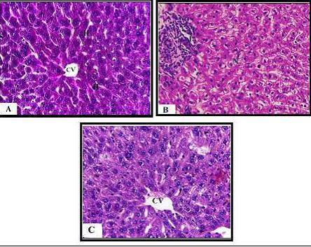
Doxorubicin, also known as Adriamycin, is a highly effective antineoplastic agent, but it is well known for its oxidative damage to various body organs, such as hepatotoxicity. The present study aimed to investigate the possible protective role of the natural antioxidant garlic oil on Doxorubicin-induced liver toxicity. Studies were performed on four groups of mice. Control group, garlic oil group, doxorubicin group for seven doses, and doxorubicin plus garlic oil group. Histological examination of liver sections revealed that doxorubicin caused pathological changes in liver cells as well as the reduction in the liver nuclear area and nuclear volume. Administration of garlic oil plus doxorubicin showed a reduction in liver damage induced by DOX.
Introduction
The liver is one of the largest and most important organs in the body and has diverse functions in metabolism, detoxification and synthesis of many biomolecules and it is the first organ affected by DOX treatment (Prasanna et al., 2020). Doxorubicin (DOX is one of the most effective anthracycline antibiotics and anti-tumour agents used to treat a variety of human malignancies, including hepatocellular carcinoma (Osama and Suzan, 2008). Doxorubicin has a greater effect on cells that are multiplying rapidly, but it can kill both healthy and cancerous cells (Saalu et al., 2009.). Doxorubicin has many side effects, including hepatotoxic, cardiotoxic, nephrotoxic, and immunosuppression (Chaudhary et al., 2016). Liver damage is a relatively common adverse effect in patients with other cancers who are treated with doxorubicin (Cainelli and Vallone, 2009). Oxidative stress is generally considered a major cause of DOX hepatotoxicity. Dox produces hydroxy radical, which destroys DNA primarily in cancerous cells. Doxorubicin can cause an imbalance of oxygen free radicals and antioxidants which causes damage to liver tissue (Mohan et al., 2014). Natural products, including plants, have been used by humans in traditional medicines to treat and alleviate different diseases and are valued for their ability to protect against all types of diseases (Wambi et al., 2009). Several natural and synthetic antioxidants have been suggested to protect against DOX-derived cardiotoxicity (Asensio-Lopez et al., 2012; El-Bakly et al., 2012). Plant phenolic compounds such as flavonoids and isoflavones have an important role in the treatment of many diseases and some of them induce a potent antioxidant and hepatoprotective effect (Seif, 2016). Garlic (Alium sativum) is a bulbous plant whose bulb has a strong taste and characteristic odor. Garlic has been well known for its medicinal use since ancient times and has been reported to have antioxidant properties in vitro (Reitz et al., 1995). Garlic is used as hypolipidemic and cardiotonic. Some garlic preparations also appear to possess hepatoprotective, immune-enhancing, anticancer, chemopreventive and antioxidant activities (Harunobu et al., 2001). In mammals, garlic has effects of hypolipidaemia, hypoglycemia, hypotriglyceridaemia and hypocholesterolaemia (Ali et al., 2000).
The aim of the present study is to investigate the potential beneficial role of garlic administration against histomorphometrical alterations induced in the liver by doxorubicin in male mice
source : The protective role of garlic oil against doxorubicin-induced hepatotoxicity in male mice
0 notes
Text
Formononetin enhances bone regeneration in a mouse model of cortical bone defect.
PMID: Br J Nutr. 2017 Jun ;117(11):1511-1522. Epub 2017 Jul 10. PMID: 28689509 Abstract Title: Formononetin, a methoxy isoflavone, enhances bone regeneration in a mouse model of cortical bone defect. Abstract: The bone regeneration and healing effect of formononetin was evaluated in a cortical bone defect model that predominantly heals by intramembranous ossification. For this study, female Balb/c mice were ovariectomised (OVx) and a drill-hole injury was generated in the midfemoral bones of all animals. Treatment with formononetin commenced the day after and continued for 21 d. Parathyroid hormone (PTH1-34) was used as a reference standard. Animals were killed at days 10 and 21. Femur bones were collected at the injury site for histomorphometry studies using microcomputed tomography (μCT) and confocal microscopy. RNA and protein were harvested from the region surrounding the drill-hole injury. For immunohistochemistry, 5 µm sections of decalcified femur bone adjoining the drill-hole site were cut. μCT analysis showed that formononetin promoted bone healing at days 10 and 21 and the healing effect observed was significantly better than in Ovx mice and equal to PTH treatment in many aspects. Formononetin also significantly enhanced bone regeneration as assessed by calcein-labelling studies. In addition, formononetin enhanced the expression of osteogenic markers at the injury site in a manner similar to PTH. Formononetin treatment also led to predominant runt-related transcription factor 2 and osteocalcin localisation at the injury site. These results support the potential of formononetin to be a bone-healing agent and are suggestive of its promising role in the fracture-repair process.
read more
0 notes
Text
Influence of Dietary Supplementation of Coated Sodium Butyrate and/or Synbiotic on Growth Performances, Caecal Fermentation, Intestinal Morphometry and Metabolic Profile of Growing Rabbits | Chapter 09 | Research and Development in Agricultural Sciences Vol. 2
Aim: The aim of the present experiment was to study the synergistic effects of dietary supplementation with coated slow released sodium butyrate (CM3000®) and a commercial synbiotic (Poultry-Star®) on the productive performance and intestinal morphometry of the growing rabbits.
Study Design: Laboratory experimental design was used in this study.
Place and Duration of Study: The study was conducted in the experimental rabbitry of Physiology Department, Faculty of Veterinary Medicine Cairo University, Egypt. The s duration of the study persists for 70 days.
Methodology: Thirty- two apparently healthy male New Zealand rabbits with average body weight of 544 ± 9 g were divided randomly into four dietary treatments at weaning (28th day of age). The control group (C) was fed on standard basal diet with no supplementation. Rabbits in the second group (T1) received the same basal diet supplemented with CM3000® 500 g/ton feed. Animals in the third group (T2) consumed the basal diet containing Poultry-Star® 500 g/ton feed. Rabbits in the fourth group (T3) were fed on the basal diet enriched with mixture of CM3000® and Poultry-Star®, 250 g/ton feed for each. Feed and water were offered ad-libitum for 70 days experimental period. Body weight and feed consumption were recorded biweekly to calculate body weight gain and feed conversion. At the end of the experimental period blood and caecal content samples were collected from all animals. Ce rtain haematological metabolic parameters namely, glucose [1] triglycerides [2], total cholesterol [3] total protein [4], albumen and urea [5]. Caecal content samples were collected at the end of the experimental period post slaughtering for determination of caecal fermentation pattern namely, pH, total short chain fatty acids [6] individual volatile fatty acids [7]and ammonia concentration [8,9]. Duodenal tissue samples were collected for histomorphometry. The results revealed that additives used improved significantly live body weight compared to the control group. Rabbits in T3 group showed the highest body weight gain. In addition, supplementation of the basal diet with a mixture of additives revealed significant increase of feed intake. The blood urea level was reduced significantly in bucks of T1. The rabbits in T3 group recorded the highest level of blood glucose. Caecal pH revealed a significant decrease in T1 and T3. The mixture of additives has positive results on the intestinal morphometry.
Conclusion: Coated butyrate and are capable of improving performance, enhancing intestinal health. Author(s) Details
Mr. Anthony Kodzo-Grey Venyo MB ChB FRCS(Ed) FRCSI FGCS Urol. LLM Department of Urology, North Manchester General Hospital, Manchester, M8 5RB, United Kingdom.
View Volume: http://bp.bookpi.org/index.php/bpi/catalog/book/132
0 notes
Text
Khasiat ulam raja yang ramai tidak tahu
http://drjamu.com.my/informasi/khasiat-ulam-raja-yang-ramai-tidak-tahu/
Khasiat ulam raja yang ramai tidak tahu
Ulam raja atau nama saintifiknya Cosmos caudatus adalah antara ulam yang paling popular di Malaysia. Ianya senang di jumpai dan senang untuk ditanam. Malah ulam raja mempunyai pelbagai khasiat yang telah dibuktikan dengan kajian saintifik. Jadi tidak hairan lah jika nenek moyang kita kerap kali mengamalkan ulam ini. Malangnya, generasi sekarang lebih terikut dengan budaya makanan barat seperti makan yang diproses. Dan ada sesetengahnya hanya mengutamakan pengambilan sayuran daripada negara luar seperti brokoli, asparagus dan lain-lain. Tanpa menyedari sayuran daripada tempat kita juga tidak kurang hebatnya (dari segi khasiat). Tapi Dr. Jamu agak terkesima, ulam raja juga berasal daripada Amerika Tengah [1]. Rupanya ulam raja juga berasal dari negara barat. hehe
Jom ketahui khasiat ulam raja
1.Tinggi antioksida
Ulam raja mengandungi pelbagai sebatian fenolik seperti proanthocyanidin, asid klorogenik, quercertin dan derivatifnya, dimana sebatian-sebatian ini telah dilaporkan sebagai antioksidan yang baik [2] . Kajian menunjukkan ulam raja mempunyai kapasiti antioksida yang sangat tinggi iaitu lebih kurang 2400 mg L-asid askorbik bersaman kapasiti bagi setiap 100 g sampel. [1]. Ulam raja juga mempunyai aktiviti antioksida yang tinggi yang diukur melalui kuasa penurun ferik sianida, hapus sisa DPPH dan pengoksidaan asid linoleik [3]. Pentingnya antioksida ini kerana ianya boleh membantu melawan penyakit yang dikaitkan dengan penuaan, kanser dan sakit jantung [4-5].
2. Untuk tulang
Kebiasanya ramai yang akan berfikir pengambilan susu baik untuk tulang. Percaya atau tidak, ulam raja juga boleh dijadikan sebagai salah satu sumber untuk menndapatkan kalsium. Malah, dilaporkan boleh merangsang pembentukan tulang [6]. Kajian juga mendapati ulam raja berpotensi to become an alternative treatment in restoring bone damage in post-menopausal women [7].
3. Peredaran darah
Sebagai rawatan tradisional untuk membantu memperbaiki peredaran darah [8].
4. Pembuka selera
Digunakan sebagai pembuka selera [9]. Dalam banyak-banyak ulam, Dr. Jamu paling menyukai rasa dan bau ulam raja. Bagi, Dr Jamu ianya lebih sedap berbanding dengan ulaman yang lain
5. Anticancer
Quercitrin iaitu salah satu sebatian yang terkandung di dalam ulam raja [10]. Ianya berpotensi bertindak sebagai agen antikaser bagi melawan penyakit kanser paru-paru [11].
6. Kecantikan kulit
Quercitrin yang bertindak sebagai antioxidant juga didapati boleh melindungi kerosakan oksidatif yang disebabkan pendedahan kepada sinaran ultraviolet B (UVB, 280-320 nm) [12]. Pendedahan kulit kepada sinaran ini akan menyebabkan kerosakan kulit seperti selaran matahari (sunburn), penuaan, dan kanser kulit [13]. Penuaan dan kerosakan kulit, selalunya dipandang serius oleh kaum wanita, jadi boleh mengamalkan ulam raja untuk kulit kekal cantik.
7. Untuk diabetes
Sebagai antidiabetic [14] dan antara ulam yang digunakan secara traditional untuk merawat pesakit diabetes (type 2) [15]. Malah kajian juga membuktikan ulam raja boleh digunakan sebagai adjuvant therapy patients with type 2-diabetes [16].
-Selamat Mencuba-
Rujukan
[1]Shui et al. 2005. Rapid screening and characterisation of antioxidants of Cosmos Caudatus using liquid chromatography coupled with mass spectrometry. Journal of Chromatography B; 127-138
[3] Andarwulan et al. 2010. Flavonoid content and antioxidant activity of vegetables from Indonesia. Food Chemistry. 121: 1231-1235
[4] Ames et al. 1993. Oxidants, antioxidants and the degenerative disease of aging. Proceeding of the National Academy of the United States of America. 90(17): 7915-7922
[5] Halliwell, B. 1996. Antioxidants in human health and disease. Annual Review of Nutrition. 16: 33-50
[6]Ismail S. 2000. Sayuran Tradisional Ulam dan Penyedap Rasa. Bangi; Universiti Kebangsaan Malaysia.
[7] Mohamed et al. 2013. The effects of Cosmos caudatus (ulam raja) on dynamic and cellular bone histomorphometry in ovariectomized rats. BMC Researh Notes, 6: 239
[8] Shui et al. 2005. Rapid screening and characterization of antioxidants of Cosmos caudatus using liquid chromatography coupled with mass spectrometry. Journal of Chromatography B. 827: 127-138.
[9] Andarwulan et al. 2012. Polyphenol, carotenoids and ascorbic acid in underutilized medicinal vegetables. Journal of functional foods. 339-347.
[10] Reihani et al. 2016. Total phenolic content and antioxidant activity of ulam raja (Cosmos caudatus) and quantification of its selected marker compounds: Effects of Extraction. International Journal of Food Properties
[11] Cincin et al. 2014. Molecular mechanisms of quercitrin-induced apoptosis in non small cell lung cancer. Archives of Medical Research. 45; 445-454
[12] Yin et al. 2013. Quercitrin protects skin from UVB-induced oxidative damage. Toxicology an Applied Pharmocology. 89-99
[13] Fisher et al. 1996. Molecular basis of sun-induced premature skin ageing and retinoid antogonism. Nature 379, 335-339.
[14] Perumal et al. 2014. Effect of Cosmos caudatus Kunth leaves on the lipid profile of a hyperlipidemia-induced animal model. Journal of Food Chemistry and Nutrition. 2(1): 43-51.
[15] Sekar et al. 2014. Ten commonly available medicinal plants in Malaysia used for the treatment of diabetes-a review. Asian Journal of Pharmeceutical and Clinical Research. 7(1)
[16]Cheng et al. 2016. Effect of Cosmos caudatus (Ulam raja) supplementation in patients with type 2 diabetes: Study protocol for a randomized controlled trial. BMC Complementary and Alternative Medicine, 16:84
0 notes
Text
Poor Glycemic Control Tied to Bone Formation in Younger Women With Type 2 Diabetes
Bone histomorphometry suggests that in premenopausal women with type 2 diabetes mellitus (T2DM) glycemic control is associated with bone remodeling, and may increase fracture risk, according to researchers in Brazil. Reuters Health Information from Medscape Medical News Headlines https://ift.tt/340Dg8q via IFTTT
0 notes
Text
In Vivo 3D Histomorphometry Quantifies Bone Apposition and Skeletal Progenitor Cell Differentiation
http://dlvr.it/QNFvvP
0 notes
Link
..
1 note
·
View note
Text
Effect of piezoelectric sutural ostectomies on accelerated bone-borne sutural expansion
Publication date: Available online 17 August 2017 Source:Journal of Oral and Maxillofacial Surgery Author(s): Akram S. Alyessary, Adrian U.J. Yap, Siti Adibah Othman, Mohammad T. Rahman, Zamri Radzi PurposeThis study investigated the effect of piezoelectric sutural ostectomies on accelerated bone-borne sutural expansion.MethodsSixteen male New Zealand white rabbits (20-24 weeks old) were randomly divided into the following four experimental groups (n=4): group 1, conventional rapid sutural expansion; group 2, accelerated sutural expansion; group 3, accelerated sutural expansion with continuous ostectomy; and group 4, accelerated sutural expansion with discontinuous ostectomy. All sutural ostectomies were performed using a piezoelectric instrument (Woodpecker DTE, DS-II., Guangxi, China) prior to expander application under anesthesia. Modified hyrax expanders were placed across the midsagittal sutures of the rabbits and secured with miniscrew implants located bilaterally in the frontal bone. The hyrax expanders were activated 0.5 mm/day for 12 days (group 1) or 2.5 mm initial expansion followed by 0.5 mm/day for 7 days (groups 2 to 4). After 6 weeks of retention, bone volume fraction, sutural separation and new bone formation were evaluated using micro-computed tomography and histomorphometry. Statistical analysis was performed using Kruskal-Wallis / Mann-Whitney U tests and Spearman’s rho correlation (p<0.05).ResultsRanking of median sutural separation was as follows: group 1 (3.05 mm), group 2 (3.97 mm), group 4 (4.78 mm) and group 3 (5.66 mm). The least and most bone formation were observed with groups 1 (63.63%) and 3 (75.93%), respectively. Spearman’s correlation showed strong, positive and significant correlation (r= 0.932, p<0.01) between the new sutural bone formation and amount of sutural separation.ConclusionPiezoelectric sutural ostectomies increase the rate of sutural separation and promotes new sutural bone formation / osteogenesis. Continuous ostectomy gave better results than discontinuous ostectomy. from # All Medicine by Alexandros G. Sfakianakis via alkiviadis.1961 on Inoreader http://ift.tt/2fQgyw4 from OtoRhinoLaryngology - Alexandros G. Sfakianakis via Alexandros G.Sfakianakis on Inoreader http://ift.tt/2uWug38
0 notes
Text
Protective action of N-acetylcysteine on sperm quality in cyclophosphamide-induced testicular toxicity.
PMID: JBRA Assist Reprod. 2019 Apr 30 ;23(2):83-90. Epub 2019 Apr 30. PMID: 30633472 Abstract Title: Protective action of N-acetylcysteine on sperm quality in cyclophosphamide-induced testicular toxicity in male Wistar rats. Abstract: BACKGROUND: Reductions in sperm quality due to free radical formation during cancer chemotherapy are well documented, hence the need for an adjunct antioxidant treatment during chemotherapy. This study was designed to investigate the effects of N-acetylcysteine on sperm quality following cyclophosphamide exposure in male Wistar rats.METHODS: wenty male Wistar rats weighing 150-170g were randomly assigned into 4 groups of five rats each, and were orally administered distilled water (Control), Cyclophosphamide (6mg/kg), N-acetylcysteine (100mg/kg) or Cyclophosphamide + N-acetylcysteine for 21 days. Sperm count, histone-protamine replacement, chromatin integrity, testicular histomorphometry and BAX Protein expression were assessed using standard procedures. The data was presented as mean± SEM and analyzed using students' t- test. A
read more
0 notes
Text
Physiological Panel of Some Feed Additives for Turkey Toms | Chapter 03 | Research and Development in Agricultural Sciences Vol. 2
Aims: To find the effect of basil and thyme (medicinal plants) and some enzymes (kemzyme and zymogen) as feed additives on some productive performance, some metabolic parameters, integrity and functionality of gastrointestinal tract.
Study Design: Forty - eight male turkey toms (strain, Big 6), 3 months old were used with an average body weight 5.5 kg. All turkeys were apparently active and healthy. The turkey toms were raised on floor covered with dry wood shaving, which was used in all partition, its thickness is about 10-15 cm and renewed every 3 days till the end of the experiment. The house was divided into 8 partitions, each partition was (240 cm X 180 cm) providing enough surface area for each turkey. In each partition there were plastic feeders and drinkers of 20 L capacity, it was provided with identifying card to record all required information. Food and water were available ad-libitum through the period of experiment. The house was well ventilated by natural windows. Light was turned off (one hour in 24 hours) as recommended by [1]. Turkey was kept under hygienic condition and as far as possible from any discomfort stimuli. Feeding Regimen; Basal ration were formulated to cover the nutrient requirements of growing turkey according to National Research Center [2]. Toms were randomly divided into eight groups (6 toms/each). The control group (C) was fed a basal diet without feed supplementation. Group(B) was supplemented with basil (3 g basil/kg diet) group (T) was fed thyme (2 g thyme/kg diet), group (TB) was fed a mixture of basil and thyme (3 g basil + 2 g thyme kg diet), group (Z) was fed zymogen (1 ml/4 liter water); and group (K) was supplemented with kemzyme (0.5 g/kg diet), group (BTK) was fed a mixture of basil, thyme and kemzyme, the last group was fed with diet supplemented with basil, thyme and zymogen (BTZ).
Place and Duration of Study: The present study was carried out in animal care center, faculty of veterinary medicine, Cairo University. During the period from March till May 2015. The experiment lasted for 10 weeks.
Methodology: Body weight gain, feed conversion ratio was measured at the end of 10th week of experiment. Blood samples were taken to measure some biochemical & hormonal parameters, at the same time some tissues for morphological and molecular studies were prepared. Tissue samples were taken from duodenum. They were stained with hematoxylin and eosin (H&E) stain for measuring the length of villi and the depth of crypts using A computerized microscopic image analyzer attached for full HD microscopic camera (Leica Microsystems, Germany was used to determine the histomorphometry parameters [3]. The other part of the collected tissues was prepared as homogenate for total DNA& RNA purification according to [4] using the QIAamp Mini Kit.
Results: Toms fed with either basil, thyme or kemzyme (BT) had significantly (p < 0.05) heaviest body gain than the control, (Z) or (BTZ). Significant increase occurred of the dressing percent (DP%) in group and (BT) compared to the groups (T, Z, BTK and BTZ).
Supplementation with (K) significantly decreases the serum total lipids than the (C) group and all supplemented groups at P < 0.05. Serum cholesterol levels of the groups (T), (BT) and (Z) recorded a significant increase than those of groups (C), (K), (BTK) and (BTZ). Meanwhile; triglyceride levels revealed significant decrease in control group at (p < 0.05) than all other groups. All experimental groups recorded no differences in both serum protein and albumin. Although there were an increased levels of serum AST of groups (T) and (Z) compared to control and other groups, meanwhile; (T) and (Z) groups revealed the lowest level of serum ALT.
Concerning the antioxidant parameters, results reviewed that (T), (BT), (BTK) and (BTZ) had a higher level of MDA activity than the (B), (K) and (Z) supplemented groups illustrate no difference versus the serum (MDA) activity of the (C) group. Serum SOD activity revealed no differences within all groups. Serum TAC recorded significant increase in all supplemented groups compared to their level in control group.
Studies of intestinal integrity; morphometry studies indicated that the villous height increased in groups (T), (Z) and (BTK) with a higher villous to crypt ratio and goblet cell numbers of group supplemented with (BTK). Moreover, the villous width revealed a significant increase in the (C) and (Z) groups. The measurement of total DNA of duodenal tissues which reflect the cell mitosis was higher in (T), (Z) and (BTK), (BTZ) at P < 0.05; the recorded results of DNA/protein of the same segment of the duodenal tissues revealed higher ratio (higher ribosomal activity) of (T), (Z), and (BTK) groups. Cell size of the duodenal tissues as indicated by (protein/DNA) was increased by (K) and decrease by (T) and (Z) supplementation at P < 0.05.
Conclusion: Supplementation of turkey rations with herbs (basil & thyme) and multienzymes (zymogen & kemzyme) either alone or in combination produce a significant improvement in turkeys’ performance. They may regulate feed intake, increase intestinal surface area for more absorption and utilization of feed; and Contain different antioxidants activity. Author(s) Details
Sohair Y. Saleh Department of Physiology, Faculty of Veterinary Medicine, Cairo University, Giza, Egypt.
Nahed S. El-Toukhy Department of Physiology, Faculty of Veterinary Medicine, Cairo University, Giza, Egypt.
Hassan. I. Abass Department of Physiology, Faculty of Veterinary Medicine, Cairo University, Giza, Egypt.
Salma I. El- Samannoudy Department of Physiology, Faculty of Veterinary Medicine, Cairo University, Giza, Egypt.
Mohamed A. Tony Department of Nutrition and Clinical Nutrition, Faculty of Veterinary Medicine, Cairo University, Giza, Egypt.
Amin M. Hassanin Department of Cytology and Histology, Faculty of Veterinary Medicine, University of Sadat City, Meonofya, Egypt.
View Volume: http://bp.bookpi.org/index.php/bpi/catalog/book/132
0 notes