#Digital Radiation Detector
Explore tagged Tumblr posts
Visit Tumblr Blog
Explore Tumblr blogs with no restrictions, modern design and the best experience.
Last Seen Tumblr Blogs


Fun Fact
BuzzFeed published a report claiming that Tumblr was utilized as a distribution channel for Russian agents to influence American voting habits during the 2016 presidential election in Feb 2018.
Text
#Digital Radiation Detector manufacturers in india#Digital Radiation Detector manufacturers#Digital Radiation Detector#Muti-Function Detectors
0 notes
Text

Digital illustration of a star shedding stellar debris as it orbits a supermassive black hole. This artist’s impression represents the center of a galaxy about 860 million light-years from Earth. Credit NASA/CXC/M.Weiss

X-ray and optical image of AT2018fyk. Credit X-ray: NASA/SAO/Kavli Inst. at MIT/D.R. Pasham; Optical: NSF/Legacy Survey/SDSS
Right on schedule: Physicists use modeling to forecast a black hole's feeding patterns with precision
The dramatic dimming of a light source ~ 860 million light-years away from Earth confirms the accuracy of a detailed model developed by a team of astrophysicists from Syracuse University, MIT and the Space Telescope Science Institute.
Powerful telescopes like NASA’s Hubble, James Webb, and Chandra X-ray Observatory provide scientists a window into deep space to probe the physics of black holes. While one might wonder how you can “see” a black hole, which famously absorbs all light, this is made possible by tidal disruption events (TDEs) - where a star is destroyed by a supermassive black hole and can fuel a “luminous accretion flare.” With luminosities thousands of billions of times brighter than the Sun, accretion events enable astrophysicists to study supermassive black holes (SMBHs) at cosmological distances.
TDEs occur when a star is violently ripped apart by a black hole’s immense gravitational field. As the star is shredded, its remnants are transformed into a stream of debris that rains back down onto the black hole to form a very hot, very bright disk of material swirling around the black hole, called an accretion disc. Scientists can study these to make direct observations of TDEs, and compare those to theoretical models to relate observations to physical properties of disrupted stars and their disrupting black holes.
A team of physicists from Syracuse University, MIT and the Space Telescope Science Institute used detailed modeling to predict the brightening and dimming of AT2018fyk, which is a repeating partial TDE, meaning the high-density core of the star survived the gravitational interaction with the SMBH, allowing it to orbit the black hole and be shredded more than once. The model predicted that AT2018fyk would “dim” in August 2023, a forecast which was confirmed when the source went dark last summer, providing evidence that their model delivers a new way to probe the physics of black holes. Their results were published in The Astrophysical Journal Letters.
A High Energy Source
Thanks to incredibly detailed extragalactic surveys, scientists are monitoring more coming and going light sources than ever before. Surveys pan entire hemispheres in search of sudden brightening or dimming of sources, which tells researchers that something has changed. Unlike the telescope in your living room that can only focus visible light, telescopes such as Chandra can detect light sources in what’s referred to as the X-ray spectrum emitted from material that is millions of degrees in temperature.
Visible light and X-rays are both forms of electromagnetic radiation, but X-rays have shorter wavelengths and more energy. Similar to the way in which your stove becomes “red hot” after you turn it on, the gas comprising a disc “glows” at different temperatures, with the hottest material closest to the black hole. However, instead of radiating its energy at optical wavelengths visible to the eye, the hottest gas in an accretion disc emits in the X-ray spectrum. These are the same X-rays used by doctors to image your bones and that can pass through soft tissue, and because of this relative transparency, the detectors used by NASA X-ray telescopes are specifically designed to detect this high-energy radiation.'
A Repeat Performance
In January 2023, a team of physicists, including Eric Coughlin, a professor at Syracuse University’s Department of Physics, Dheeraj R. “DJ” Pasham, a research scientist at MIT, and Thomas Wevers, a Fellow at the Space Telescope Science Institute, published a paper in The Astrophysical Journal Letters that proposed a detailed model for a repeating partial TDE. Their results were the first to map a star’s surprising return orbit about a supermassive black hole – revealing new information about one of the cosmos’ most extreme environments.
The team based their study on a TDE known as AT2018fyk (AT stands for “Astrophysical Transient”), where a star was proposed to be captured by a SMBH through an exchange process known as “Hills capture.” Originally part of a binary system (two stars that orbit one another under their mutual gravitational attraction), one of the stars was hypothesized to have been captured by the gravitational field of the black hole and the other (non-captured) star was ejected from the center of the galaxy at speeds comparable to ~ 1000 km/s.
Once bound to the SMBH, the star powering the emission from AT2018fyk has been repeatedly stripped of its outer envelope each time it passes through its point of closest approach with the black hole. The stripped outer layers of the star form the bright accretion disk, which researchers can study using X-Ray and Ultraviolet /Optical telescopes that observe light from distant galaxies.
While TDEs are usually “once-and-done” because the extreme gravitational field of the SMBH destroys the star, meaning that the SMBH fades back into darkness following the accretion flare, AT2018fyk offered the unique opportunity to probe a repeating partial TDE.
The research team has used a trio of telescopes to make the initial and follow-up detections: Swift and Chandra, both operated by NASA, and XMM-Newton, which is a European mission. First observed in 2018, AT2018fyk is ~ 870 million light years away, meaning that because of the time it takes light to travel, it happened in “real time” ~ 870 million years ago.
The team used detailed modeling to forecast that the light source would abruptly disappear around August 2023 and brighten again when the freshly stripped material accretes onto the black hole in 2025.
Model Validation
Confirming the accuracy of their model, the team reported an X-ray drop in flux over a span of two months, starting on August 14, 2023. This sudden change can be interpreted as the second emission shutoff.
“The observed emission shutoff shows that our model and assumptions are viable, and suggests that we are really seeing a star being slowly devoured by a distant and very massive black hole,” says Coughlin. “In our paper last year, we used constraints from the initial outburst, dimming and rebrightening to predict that AT2018fyk should display a sudden and rapid dimming in August of 2023, if the star survived the second encounter that fueled the second brightening.”
The fact that the system displayed this predicted shutoff therefore implies several distinctions about the star and the black hole:
the star survived its second encounter with the black hole;
the rate of return of stripped debris to the black hole is tightly coupled to the brightness of AT2018fyk;
and the orbital period of the star about the black hole is ~ 1300 days, or about 3.5 years.
The second cutoff implies that another rebrightening should happen between May and August of 2025, and if the star survived the second encounter, a third shutoff is predicted to occur between January and July of 2027.
As for whether we can count on seeing a rebrightening in 2025, Coughlin says the detection of a second cutoff implies that the star has had more mass freshly stripped, which should return to the black hole to produce a third brightening.
“The only uncertainty is in the peak of the emission,” he says. “The second re-brightened peak was considerably dimmer than the first, and it is, unfortunately, possible that the third outburst will be dimmer still. This is the only thing that would limit the detectability of this third outburst.”
Coughlin notes that this model signifies an exciting new way to study the incredibly rare occurrences of repeating partial TDEs, which are believed to take place once every million years in a given galaxy. To date, he says scientists have encountered only four to five systems that display this behavior.
“With the advent of improved detection technology uncovering more repeating partial TDEs, we anticipate that this model will be an essential tool for scientists in identifying these discoveries,” he says.
8 notes
·
View notes
Text
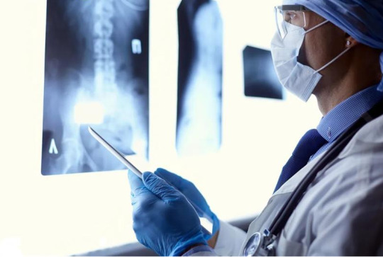
Why a High-Quality X-Ray Image Can Make or Break a Diagnosis or Surgery
In modern healthcare, an X-ray image is like a doctor’s second pair of eyes. From diagnosing bone fractures to guiding high-precision spinal surgeries, a clear, properly positioned X-ray image is the cornerstone of accurate clinical decision-making.
So, what makes a truly “high-quality” X-ray? Let’s explore the answer through the lens of Perlove Medical’s professional imaging technology.
1. Proper Patient Positioning = Valuable Imaging
Some patients present previous X-rays that are tilted, misaligned, or poorly centered — making it hard for doctors to accurately assess the problem. Poor positioning can lead to misjudgments or missed diagnoses.
Perlove Medical’s digital radiography systems (such as the PLX5100 and PLX5500) feature precision positioning mechanics and smart interfaces that help radiographers quickly align the imaging region, ensuring scientifically accurate patient posture and image angles — so doctors don’t have to “guess.”
2. Clear Image Quality Builds Clinical Confidence
Image sharpness directly determines whether the doctor can “see” key anatomical structures. Perlove Medical’s high-end dynamic DR and mobile C-arm systems use advanced flat panel detectors and proprietary image processing algorithms to deliver high-resolution, high-contrast, low-noise images. This clarity reveals fine details — from fracture lines to soft tissue contours.
For example, during spinal procedures, surgeons rely on a clear visualization of the “cat’s eye” sign — a critical X-ray landmark that helps ensure screws are precisely placed within the pedicle. Perlove’s mobile C-arm systems (like the PLX118F and PLX7500) are tailored for such demanding tasks, providing high frame rates and instant imaging that enhance intraoperative precision.
📌 Knowledge Tip: The “cat’s eye” refers to the pedicle shadow in anteroposterior (AP) X-ray views, resembling a feline pupil. It’s a key visual indicator for proper pedicle screw placement.
3. Proper Exposure — Not Too Bright, Not Too Blurry
Overexposed or blurry images can obscure diagnostic details. Perlove Medical’s DR systems are equipped with Automatic Exposure Control (AEC) technology, which intelligently adjusts radiation levels based on tissue density — protecting the patient while ensuring image clarity.
Meanwhile, intelligent image optimization enhances contrast and detail across regions of interest, including bone, soft tissue, and metallic implants — making Perlove’s systems ideal for orthopedics, emergency care, neurosurgery, and beyond.
4. 2D + 3D Imaging = Safer, Smarter Surgeries
Traditional 2D X-rays require surgeons to rely on experience to judge depth and alignment — leaving room for error. Perlove Medical’s advanced 3D C-arm (such as the PLX7200) supports real-time 3D reconstruction, upgrading standard fluoroscopy to a 3D view that reveals bone structure and implant paths in detail.
Surgeons can directly visualize whether a screw is safely inside the pedicle or deviated from its intended path — enabling accurate intraoperative adjustments, increasing safety, and boosting the success rate of complex surgeries.
🎯 Conclusion: Clinical excellence begins with clear imaging — and behind every high-quality X-ray is a high-performance device. Perlove Medical is committed to empowering medical professionals with advanced imaging solutions that help them see better, decide faster, and operate with greater precision.
📍 For more product information and clinical case demonstrations, please visit the Perlove Medical official website or contact an authorized distributor.

1 note
·
View note
Text
Digital alpha-electron discrimination with a silicon detector and its application as an auxiliary detector in the alpha TOF spectrometer
Alpha Time-Of-Flight (A-TOF) spectroscopy is commonly used to measure alpha particles in high-energy resolution. To minimize interference from background radiation and prevent random TOF registrations during long-time measurements, specific strategies are necessary.
Global Particle Physics Excellence Awards
website url: physicistparticle.com/
Nomination link: https://physicistparticle.com/award-nomination/?ecategory=Awards&rcategory=Awardee
For Enquiry: [email protected]
#sciencefather#SiliconDetector#AlphaParticleDetection#TOFSpectrometry#DigitalSignalProcessing#NuclearInstrumentation#RadiationDetection#ParticlePhysics
1 note
·
View note
Text

"With technology playing such a big role in today’s business world, what tech-related skills do you think are the most important for aspiring entrepreneurs to succeed?" I approached a fellow student, a tech-savvy girl who radiated quiet confidence. When I asked her about the role of technology in entrepreneurship, she took a moment to reflect. It was clear she had thought deeply about this topic before.
"Honestly," she began, "the most important skill is knowing how to leverage technology to scale your business. It's not enough to just have a great idea anymore. You need to understand the tools and platforms that can amplify that idea." She talked about how technology drives everything now — from digital marketing to data analytics, even automation. "You have to be comfortable with tech, and not just the basics. I’m talking about using social media strategically, analyzing customer data, and automating repetitive processes."
I nodded in agreement, but then she said something that really made me sit up. "You know, entrepreneurs today should also be thinking about how technology can solve real-world problems, not just make businesses more efficient." That’s when she gave me an idea that completely shifted my mindset.
"What if there was a way to create an automatic earthquake detector for our campus buildings? Something that could detect tremors and instantly trigger an alarm without anyone having to manually push a button?" Her eyes lit up as she explained further. "Think about it — a system that could send alerts through the building's intercom, text notifications to everyone’s phones, and automatically alert emergency services, all without human intervention."
It was a brilliant concept — and it immediately got my gears turning. I could see the potential for something like this, especially in earthquake-prone regions. It wasn’t just about making businesses more profitable; this was about saving lives with technology. "An automatic earthquake detector," I repeated, imagining the possibilities.
Her idea opened my eyes to the broader role technology can play, not just in entrepreneurship, but in society. Entrepreneurs who can harness technology to address real-world challenges, like safety and disaster preparedness, have the potential to make a lasting impact.
She continued by stressing how important adaptability is in tech-driven industries. "The skills you learn today could be outdated in a few years, so you have to be a lifelong learner. And don’t be afraid of failure — it’s part of the process when you’re innovating with new technology."
We talked about specific technologies that are shaping the entrepreneurial landscape, like data analytics and cloud computing. But the real takeaway from our conversation was the power of combining technical skills with creative thinking. Her idea for an earthquake detector wasn’t just a reminder of how crucial technology is — it showed me how entrepreneurs can use tech to create real, meaningful change.
As I walked away from the conversation, my mind was racing with possibilities. It was clear that technology is no longer just an accessory to entrepreneurship — it's a driving force. And entrepreneurs, like this student, who can combine technical know-how with visionary thinking, are the ones who will truly make a difference in the future.
#TechInEntrepreneurship#InnovationInAction#SmartTechSolutions#EntrepreneurMindset#TechForSafety#FutureOfBusiness#AIInnovation#DisasterPreparedness#AutomationInBusiness#CreativeEntrepreneurship#LifelongLearning#TechSavvy
2 notes
·
View notes
Text
“But no one actually ‘looks’ through [modern telescopes]. Margaret Huggins lamented the shift from gazing at the heavens to squinting at tiny patches of light. Now we’ve gone much, much further. In today’s astronomy, photons of light from the sky, along with the celestial secrets they contain, are picked up by electronic detectors, converted into digital data and crunched through impossibly complex equations by some of the most powerful computers on the planet. In 2016, bricklayer-turned-astronomer Gary Fildes described visiting Chile’s Very Large Telescope (VLT) in his best-selling book An Astronomer’s Tale. Incorporating four mirrors, each 27 feet wide, the VLT collects visible and infrared radiation and can distinguish points in the sky separated by less than a millionth of a degree. Here, at the forefront of today’s attempts to understand the stars, Fildes was struck by the sight of scientists hard at work in control rooms, eyes glued not to their telescopes but to banks of screens: ‘They didn’t look as if they had seen the real sky for days.’”
- The Human Cosmos: Civilization and the Stars by Jo Marchant
#brot posts#astro posting#GOD this puts to words something i really felt#as someone who fell in love with the idea of astronomy as this awe-filled wonder of the vast universe#and then going to college and sitting in a fucking dark classroom at the brink of dawn fucking 8am and doing nothing but MATH !!!!#like - theres no judgment here#very very obviously we need these technologies and math techniques to truly understand astronomy#but like the whole thesis to this book (so far? im thinking) is that like#in doing so - you lose something fundamental#astronomy is one of if not theee oldest sciences known to humanity#but the way it was practiced for millennia upon millennia of human history is so incredibly different from how we practice it now#i got a whole ass Bachelors of Science in Astronomy and never once was i required to actually look at the night sky .#and i dont think this same phenomenon exists in other fields of science#like as time goes on we ofc learn more and theres a certain level of abstraction as you get more separated from the immediate knowledge#afforded by your immediate senses#but the level of abstraction for astronomy is just. not really seen as much or as bad in other fields? imo?#anyway i remember a while ago saying that as i got further through my degree the less magical space felt to me#compared to when i was younger and knew nothing at all#and i said yeah its nice to /know/ things now but i still miss that magic when everything was new exciting and real#but you lose something. that magic. that soul. when your astronomg experience is not actually stargazing#but instead sitting in a room doing math on paper or doing nothing but staring at a computer screen
6 notes
·
View notes
Text
Non-contact Infrared Temperature Sensor Market - Trends, Growth, including COVID19 Impact, Forecast
Global Non-contact Infrared Temperature Sensor Market Research Report 2025(Status and Outlook)
The global Non-contact Infrared Temperature Sensor Market size was valued at US$ 847.2 million in 2024 and is projected to reach US$ 1.42 billion by 2032, at a CAGR of 7.38% during the forecast period 2025-2032.
Non-contact infrared temperature sensors detect thermal radiation emitted by objects to measure surface temperature without physical contact. These sensors utilize thermopile technology that converts infrared energy into electrical signals, with key components including optical systems, detectors, and signal processing circuits. The technology finds applications across medical devices, industrial processes, automotive systems, and smart home appliances.
The market growth is driven by increasing demand for contactless temperature measurement in healthcare (especially post-pandemic), industrial automation trends, and stringent safety regulations. Recent technological advancements in sensor accuracy (±0.1°C tolerance) and miniaturization (chip-scale packaging) are expanding application possibilities. Key players like Melexis and TE Connectivity have introduced innovative MEMS-based sensors with digital outputs and IoT compatibility, while companies such as Excelitas Technologies and Hamamatsu Photonics continue to dominate the high-precision industrial sensor segment.
Our comprehensive Market report is ready with the latest trends, growth opportunities, and strategic analysis.https://semiconductorinsight.com/download-sample-report/?product_id=95914
Segment Analysis:
By Type
Through Hole Thermopile Infrared Sensor Segment Maintains Strong Market Share Due to Wide Industrial Applications
The market is segmented based on type into:
Through Hole Thermopile Infrared Sensor
SMD Thermopile Infrared Sensor
Other advanced variants
By Application
IoT Smart Home Segment Shows Rapid Growth With Increasing Adoption of Automation Technologies
The market is segmented based on application into:
IoT Smart Home
Industrial Use
Healthcare
Automotive
Others
By Technology
Advanced MEMS-based Sensors Gain Traction Due to Miniaturization and Higher Accuracy
The market is segmented based on technology into:
MEMS-based sensors
Thin film thermopiles
Traditional thermopile sensors
By End-User Industry
Manufacturing Sector Remains Key Consumer With Growing Automation Needs
The market is segmented based on end-user industry into:
Manufacturing
Healthcare
Consumer Electronics
Automotive
Others
Regional Analysis: Global Non-contact Infrared Temperature Sensor Market
North America The North American market for non-contact infrared temperature sensors is driven by stringent healthcare and industrial safety regulations, particularly in the U.S., where demand surged during the COVID-19 pandemic. The FDA-approved use of these sensors in medical diagnostics and their adoption in smart building automation systems contribute to steady growth. The region’s focus on Industry 4.0 integration and IoT applications in manufacturing further accelerates demand. Major players like Excelitas and TE Connectivity dominate the market, leveraging advanced thermopile sensor technology. However, high production costs and competition from alternative sensing technologies pose challenges.
Europe Europe maintains a strong position in the market, supported by strict workplace safety standards under EU directives and the widespread adoption of energy-efficient building management systems. Germany and the UK lead in industrial applications, while France emphasizes healthcare integrations. The region’s emphasis on automotive manufacturing (e.g., thermal imaging for EVs) and intelligent HVAC systems drives innovation. However, GDPR compliance for data collection via sensors adds complexity. Leading manufacturers like Melexis and Heimann Sensor have intensified R&D to meet these regulatory and technical demands.
Asia-Pacific As the fastest-growing regional market, China accounts for over 40% of global production due to its electronics manufacturing ecosystem. The pandemic-driven demand for fever screening devices led to exponential growth, with domestic players like Winson and Senba Sensing expanding capacity. India and Southeast Asia are emerging markets, fueled by smart city initiatives and increasing industrial automation. While cost-effective SMD thermopile sensors dominate, Japan and South Korea focus on high-accuracy applications in robotics and automotive sectors. The region’s challenge lies in balancing affordability with precision requirements.
South America Market growth in South America has been slower due to economic volatility and limited local manufacturing capabilities. Brazil represents the largest market, primarily for industrial equipment maintenance applications, while Argentina sees growing use in food processing temperature monitoring. The lack of standardized regulations compared to North America or Europe results in fragmented adoption patterns. Nonetheless, increasing foreign investments in mining and oil/gas sectors are creating new opportunities for infrared temperature sensing solutions in hazardous environments.
Middle East & Africa The MEA market shows potential with rising smart infrastructure projects in the UAE and Saudi Arabia, where non-contact sensors are deployed in building automation and oil refinery safety systems. Africa’s adoption is constrained by limited technical expertise and infrastructure, though South Africa and Nigeria are emerging as key markets for industrial and medical applications. The region benefits from government initiatives to modernize healthcare facilities but struggles with reliance on imports due to underdeveloped local manufacturing ecosystems.
List of Key Non-Contact Infrared Temperature Sensor Manufacturers
Excelitas Technologies Corp. (U.S.)
Orisystech (South Korea)
Heimann Sensor GmbH (Germany)
Melexis NV (Belgium)
Amphenol Advanced Sensors (U.S.)
TE Connectivity Ltd. (Switzerland)
Semitec Corporation (Japan)
Hamamatsu Photonic KK (Japan)
Nicera Co., Ltd. (Japan)
KODENSHI Corporation (Japan)
Winson Electronics (Taiwan)
Senba Sensing Technology (China)
Sunshine Technologies (China)
San-U (South Korea)
The global non-contact infrared temperature sensor market is witnessing substantial growth due to increasing adoption across healthcare facilities. These sensors have become indispensable tools for mass fever screening, particularly in hospitals, airports, and public spaces – a trend accelerated by recent global health crises. Medical-grade infrared thermometers now account for over 35% of total market revenue, with demand consistently growing at 12% annually. Their ability to provide rapid, hygienic temperature readings without physical contact makes them ideal for infection control protocols. Furthermore, technological advancements have improved accuracy to ±0.2°C, making them suitable for critical medical applications.
Industrial sectors are increasingly incorporating infrared temperature sensors into automation systems, driving market growth. Manufacturing facilities utilize these sensors for equipment monitoring, predictive maintenance, and quality control processes where traditional contact sensors are impractical. The Industry 4.0 revolution has particularly boosted adoption, with smart factories deploying these sensors across production lines. Recent data shows that industrial applications now represent 28% of the market share, growing at 9% year-over-year. Their durability in harsh environments and ability to measure moving objects make them invaluable for modern manufacturing operations.
The proliferation of IoT-enabled devices is further amplifying demand, as infrared sensors can be seamlessly integrated into wireless monitoring systems. This integration reduces downtime by enabling real-time temperature tracking of critical machinery components.
The rapid expansion of smart home ecosystems offers significant opportunities for non-contact infrared temperature sensor manufacturers. Integration with home automation systems for climate control, appliance monitoring, and energy management represents a growing market segment projected to grow at 18% CAGR over the next five years. Advanced sensors capable of multi-point temperature mapping are particularly in demand for next-generation HVAC systems.
Additionally, the development of compact, low-power consumption sensors compatible with IoT platforms enables new use cases in residential security and eldercare applications. Major technology companies are actively seeking partnerships with sensor manufacturers to develop integrated solutions for the connected home market.
The market is highly fragmented, with a mix of global and regional players competing for market share. To Learn More About the Global Trends Impacting the Future of Top 10 Companies https://semiconductorinsight.com/download-sample-report/?product_id=95914
Key Questions Answered by the Non-contact Infrared Temperature Sensor Market Report:
What is the current market size of Global Non-contact Infrared Temperature Sensor Market?
Which key companies operate in Global Non-contact Infrared Temperature Sensor Market?
What are the key growth drivers?
Which region dominates the market?
What are the emerging trends?
Browse More Reports:
CONTACT US:
City vista, 203A, Fountain Road, Ashoka Nagar, Kharadi, Pune, Maharashtra 411014
[+91 8087992013]
0 notes
Text
Everything You Need to Know about Getting an X-Ray

X-rays rank among the most commonly performed medical procedures in the United States. Per the Journal of the American Medical Association, the rate of radiography in the United States was 783 examinations per 1,000 people in 2024, accounting for more than 55 percent of all medical imaging procedures. Although X-ray procedures are fairly standard, it is normal for people to feel apprehensive before any medical procedure. To calm one's mind, it can be helpful to learn about what to expect from an X-ray appointment.
X-rays use electromagnetic radiation to produce images of tissues and various structures inside the body. By simultaneously projecting X-rays through a human body and an X-ray detector, medical professionals can produce an image made up of "shadows" and shapes that represent objects within the body. X-ray images, or radiographs, are often produced using photographic film, but different X-ray detectors produce different types of images, including digital images.
X-ray technology can be used to produce radiographs of virtually every part of the body and can help support diagnostics for a wide range of injuries and illnesses, from common dental problems to chronic diseases. X-rays can be used to detect or confirm broken or fractured bones, scoliosis, malignant and benign tumors, and health conditions impacting the lungs, from pneumonia to lung cancer. Radiographs can also be used to diagnose issues of the cardiovascular system, including heart failure, the leading cause of death in the US, according to the Centers for Disease Control and Prevention.
Individuals should feel comfortable discussing all aspects of an X-ray procedure with their health care provider. While physicians may provide patients with specific insight, there are a few common steps individuals can take to prepare themselves for an X-ray.
In most cases, X-ray preparations are relatively straightforward. Unlike with certain imaging procedures, individuals do not need to abstain from eating or drinking in the hours leading up to an X-ray. That said, certain medications can impact radiographs produced using contrast agents; doctors will inform patients if they need to suspend their medications and for how long prior to the procedure.
While primary care providers should already have this information, pregnant women should inform X-ray technicians of their condition. X-rays are typically not recommended for pregnant women unless the procedure is considered vital to the patient's overall health.
Finally, while X-ray procedures are usually relatively quick, patients are advised to dress comfortably. Loose clothing is best. Medical professionals advise patients to leave jewelry and clothing with metal features, including zippers, at home; otherwise, they may need to be removed.
Patients have to do very little once the procedure begins, other than remaining still for the duration. Individuals may be standing, seated, or lying down during an X-ray. The radiographer will aim the machine at the correct location on the body and operate the machine from another room. The process of producing a radiograph takes fractions of a second, and individuals do not feel any pain, discomfort, or any sensation to speak of during the process. The entire procedure may take several minutes if the radiographer needs to capture images from multiple angles.
0 notes
Text
0 notes
Text
0 notes
Text
Portable X ray veterinary
Technological Origins and Development
The ability to peer inside a living body transformed human medicine, and veterinary practice was no exception. While traditional fixed X-ray systems became staples in clinics, the need for imaging large, immobile, or distressed animals where they are drove the development of portable veterinary X-ray systems. Their origins lie in adapting early, cumbersome portable human units, but significant miniaturization, power efficiency improvements, and crucially, the transition from film to Computed Radiography (CR) and then Digital Radiography (DR) panels revolutionized the field. The introduction of lightweight, battery-powered generators coupled with durable, highly sensitive digital detectors in the late 1990s and early 2000s truly unlocked portability. Modern systems are remarkably compact, often featuring wireless DR panels, enabling true point-of-care imaging.
Clinical Applications and Advantages
The clinical impact of portable veterinary X-ray is profound.
Its core advantage is mobility: imaging occurs stall-side, barn-side, in the field, or even in the comfort of a pet's home. This drastically reduces stress for patients, especially large animals like horses or fearful pets, eliminating risky transportation or complex sedation.
It significantly improves workflow efficiency——a fractured leg on a cow or a respiratory emergency in a critical canine can be diagnosed immediately. Portability enables essential imaging in emergency situations (trauma, colic) and during surgical procedures (confirmation of implant placement, locating foreign bodies).
For practitioners, the instant digital images allow for rapid assessment, better client communication by showing results on-site, and efficient sharing with specialists for consultations.
The reduced need for heavy shielding in multiple rooms (compared to fixed units) also offers cost and space savings for clinics.

Future Directions
The future of portable veterinary X-ray is bright and interconnected. Enhanced integration with practice management software and cloud storage for seamless access to patient records and images is key. Artificial Intelligence (AI) is poised to play a significant role, offering automated preliminary analysis (e.g., fracture detection, lung pattern recognition) to augment the veterinarian's diagnosis, especially valuable in field conditions. We can expect continued improvements in image quality and detector durability alongside longer battery life and potentially lighter, even more ergonomic designs. Connectivity will advance, enabling real-time remote specialist viewing during procedures.
Challenges and Industry Trends
Initial investment costs for high-quality DR systems can be significant, though long-term savings on film/chemicals and efficiency gains offset this.
Ensuring consistent radiation safety for personnel in diverse, often uncontrolled environments requires rigorous protocols, training, and appropriate personal protective equipment (PPE).
Image quality optimization can be trickier in suboptimal lighting or with uncooperative patients compared to a controlled radiology room. Furthermore, navigating complex and varying regulatory requirements across different regions adds an administrative layer.
Industry trends clearly indicate robust growth. The rising demand for advanced pet care, including from mobile veterinary services and specialty practices, is a major driver. Increased adoption in large animal and equine medicine continues, while even small animal clinics appreciate the flexibility for critical care, dentistry, and exotics. Technological convergence, blending portability with advanced capabilities like fluoroscopy or higher power for challenging anatomies, is emerging. The focus is also shifting towards user-friendly interfaces and comprehensive training solutions to maximize the technology's potential and ensure safety.
Conclusion
Portable veterinary X-ray technology is far more than just a convenient alternative to fixed systems; it's a transformative tool that redefines the point of care. By bringing diagnostic imaging directly to the patient ——whether in a stable, a pasture, an exam room, or an emergency site ——it enhances animal welfare, improves diagnostic speed and accuracy, streamlines workflows, and expands the capabilities of veterinary professionals in diverse settings. As technology continues to evolve, becoming smarter, more connected, and more accessible, portable X-ray will remain at the forefront of delivering advanced, compassionate, and efficient veterinary medicine, ensuring better outcomes for animals wherever they may be.
0 notes
Text
0 notes
Text
Dental Caries Detectors Market Size, Market Growth & Innovations 2032
Dental Caries Detectors Market Overview The global Dental Caries Detectors Market is witnessing significant growth, driven by increasing awareness around oral health, early diagnosis technologies, and rising dental tourism. As of 2024, the market is valued at approximately USD 350 million and is projected to grow at a CAGR of 7.8% from 2025 to 2032, potentially surpassing USD 600 million by the end of the forecast period. The growing prevalence of dental caries, particularly among aging populations and children, fuels the demand for early diagnostic tools. Advancements in intraoral cameras, laser fluorescence devices, and near-infrared transillumination (NIRI) are transforming the way dentists detect cavities at non-visible stages. Dental Caries Detectors Market Dynamics The market is propelled by several strong growth drivers. Increasing healthcare expenditure on preventive dental care, rising adoption of minimally invasive diagnostic technologies, and expanding access to dental clinics are key contributors. The surge in demand for chairside caries detection systems and digital imaging solutions enhances patient outcomes while improving workflow efficiency. However, restraints such as high initial investment costs, limited reimbursement policies, and lack of skilled personnel in emerging regions restrict market expansion. Despite these hurdles, opportunities abound in untapped markets, particularly in Asia-Pacific and Latin America, where awareness of dental hygiene is growing. Additionally, the integration of artificial intelligence (AI) in diagnostic tools, along with regulatory support for innovative dental technologies, is expected to bolster market performance. Sustainability also plays a growing role, with eco-friendly, radiation-free detection methods gaining popularity among clinics aiming to minimize their environmental impact. Download Full PDF Sample Copy of Dental Caries Detectors Market Report @ https://www.verifiedmarketresearch.com/download-sample?rid=249181&utm_source=PR-News&utm_medium=365 Dental Caries Detectors Market Trends and Innovations Several notable trends are shaping the future of the Dental Caries Detectors Market. One major trend is the digitization of diagnostic dentistry. Devices utilizing AI and machine learning algorithms are enabling early, accurate, and real-time caries detection. Manufacturers are investing in compact, wireless, and ergonomic devices to enhance usability for dental professionals. Integration with cloud-based data platforms further enables remote diagnostics and seamless data sharing. Emerging product innovations include dual-mode systems combining fluorescence imaging with NIRI, which significantly improve diagnostic accuracy. Companies are also focusing on collaborative ventures with dental universities and research institutes to develop user-friendly diagnostic systems with enhanced sensitivity and specificity. Dental Caries Detectors Market Challenges and Solutions The market faces several challenges, including pricing pressures due to intense competition among device manufacturers and economic disparities across regions. Supply chain disruptions, especially those witnessed during global crises, continue to hinder timely product delivery and inventory management. Regulatory hurdles related to device approval processes also slow market entry for innovative technologies. To overcome these issues, manufacturers are adopting localized production strategies, establishing partnerships with regional distributors, and investing in scalable, modular technologies. In parallel, governments and regulatory bodies are working toward faster certification processes for dental diagnostic devices, especially those addressing public health concerns. Dental Caries Detectors Market Future Outlook The Dental Caries Detectors Market is poised for robust expansion over the next decade. Increasing patient demand for preventive dental care, coupled with continued innovation in detection technologies, will drive long-term growth.
The integration of AI-powered diagnostic systems with electronic health records and mobile dental platforms is expected to transform clinical workflows and improve patient outcomes. Rising investments in dental infrastructure, growing dental insurance penetration, and strategic market expansions by global players will continue to shape the competitive landscape. As a result, the market will transition from basic visual-tactile methods to digitally driven, evidence-based caries detection, reflecting a paradigm shift in modern dentistry. Key Players in the Dental Caries Detectors Market Dental Caries Detectors Market are renowned for their innovative approach, blending advanced technology with traditional expertise. Major players focus on high-quality production standards, often emphasizing sustainability and energy efficiency. These companies dominate both domestic and international markets through continuous product development, strategic partnerships, and cutting-edge research. Leading manufacturers prioritize consumer demands and evolving trends, ensuring compliance with regulatory standards. Their competitive edge is often maintained through robust R&D investments and a strong focus on exporting premium products globally. Dentsply Sirona Kavo Dental Quantum Dental Technologies Inc. Acteon Group Hu-Friedy Mfg. Co. AdDent Inc. DentLight Inc. Air Techniques Inc. Get Discount On The Purchase Of This Report @ https://www.verifiedmarketresearch.com/ask-for-discount?rid=249181&utm_source=PR-News&utm_medium=365 Dental Caries Detectors Market Segments Analysis and Regional Economic Significance The Dental Caries Detectors Market is segmented based on key parameters such as product type, application, end-user, and geography. Product segmentation highlights diverse offerings catering to specific industry needs, while application-based segmentation emphasizes varied usage across sectors. End-user segmentation identifies target industries driving demand, including healthcare, manufacturing, and consumer goods. These segments collectively offer valuable insights into market dynamics, enabling businesses to tailor strategies, enhance market positioning, and capitalize on emerging opportunities. The Dental Caries Detectors Market showcases significant regional diversity, with key markets spread across North America, Europe, Asia-Pacific, Latin America, and the Middle East & Africa. Each region contributes uniquely, driven by factors such as technological advancements, resource availability, regulatory frameworks, and consumer demand. Dental Caries Detectors Market, By Product • Laser Fluorescent Caries Detector• Fiber Optic Trans-illumination Caries Detector Dental Caries Detectors Market, By End-User • Hospitals• Dental Clinics• Ambulatory Surgical Centers Dental Caries Detectors Market By Geography • North America• Europe• Asia Pacific• Latin America• Middle East and Africa For More Information or Query, Visit @ https://www.verifiedmarketresearch.com/product/dental-caries-detectors-market/ About Us: Verified Market Research Verified Market Research is a leading Global Research and Consulting firm servicing over 5000+ global clients. We provide advanced analytical research solutions while offering information-enriched research studies. We also offer insights into strategic and growth analyses and data necessary to achieve corporate goals and critical revenue decisions. Our 250 Analysts and SMEs offer a high level of expertise in data collection and governance using industrial techniques to collect and analyze data on more than 25,000 high-impact and niche markets. Our analysts are trained to combine modern data collection techniques, superior research methodology, expertise, and years of collective experience to produce informative and accurate research. Contact us: Mr. Edwyne Fernandes US: +1 (650)-781-4080 US Toll-Free: +1 (800)-782-1768 Website: https://www.verifiedmarketresearch.com/ Top Trending Reports https://www.verifiedmarketresearch.com/ko/product/us-cable-market/
https://www.verifiedmarketresearch.com/ko/product/japan-north-america-and-europe-tanker-trucks-market/ https://www.verifiedmarketresearch.com/ko/product/fosfomycin-market/ https://www.verifiedmarketresearch.com/ko/product/us-and-canada-wood-plastic-composites-wpc-market/ https://www.verifiedmarketresearch.com/ko/product/middle-east-and-africa-sea-air-logistics-market/
0 notes
Text
C-Arms Market to Grow at 9.8% CAGR | USD 3.05 B in 2021
Market Size & Forecast
The C-Arms Market was valued at USD 3,054.70 million in 2021 and is expected to grow at a compound annual growth rate (CAGR) of 9.80% over the forecast period 2024–2031. This significant growth is driven by the rising demand for minimally invasive surgical procedures, technological advancements in imaging, and increased healthcare investments, particularly in developed European countries.
Introduction & Definition

Market Drivers & Restraints
Drivers:
Growth of minimally invasive procedures, offering reduced recovery time, lower risk, and optimized outcomes
Technological upgrades, such as 3D imaging, AI-enabled systems, and compact mobile units for ambulatory centers
Rising prevalence of chronic diseases—especially cardiovascular and orthopedic conditions—fuels procedural volume
Restraints:
High acquisition and maintenance costs, particularly for advanced flat-panel and robotic C-Arms
Preference for refurbished units in emerging markets due to limited budgets. To get a free sample report, click on https://www.datamintelligence.com/download-sample/c-arms-market
Segmentation Analysis
By Type: Mobile C-Arms—from full-sized to mini—dominate, thanks to their flexibility and utility in a wide range of procedures
By Detector: Flat-panel detectors capture the largest share due to superior image quality, speed, and lower radiation exposure
By Application: Cardiology remains the leading segment (e.g., angiography, structural heart interventions), closely followed by orthopedics & trauma; oncology and gastroenterology are also showing significant growth
By End User: Hospitals lead adoption, followed by diagnostic centers and specialty clinics, supported by expanding outpatient imaging infrastructure To get the unlimited market intelligence, subscribe to https://www.datamintelligence.com/reports-subscription
Geographical Insights
Germany & UK: Early adopters with strong hospital networks and robust reimbursement frameworks for imaging procedures
France, Italy & Spain: Moderate growth driven by healthcare modernization and public funding for surgical imaging
Nordics & Benelux: Adopting advanced systems early, particularly in AI-based radiology and 3D imaging
Central & Eastern Europe: Slower uptake due to cost concerns but gradually moving toward mobile and digital upgrades
Recent Trends & Industry Highlights
AI-powered imaging is becoming mainstream, improving image clarity, dosage reduction, and workflow automation
3D mapping and hybrid systems enhance surgical precision—developers like Philips and Siemens are leading in this space
Robotic integration and linkages with navigation systems are increasing procedural efficiency and enabling advanced endovascular and orthopedic workflows
Competitive Landscape
Key manufacturers operating across Europe include:
GE HealthCare
Philips
Siemens Healthineers
Canon Medical Systems
Hologic
These companies are focusing on product innovation—especially flat-panel detectors, AI-enabled workflow, and 3D imaging—as well as strategic partnerships and aftermarket services to strengthen regional presence.
Key Developments
Philips launched new AI-enabled C-Arm units, reducing radiation and improving procedural efficiency
Siemens, GE, and Canon introduced upgraded mobile systems with 3D imaging capabilities
Ziehm Imaging introduced new-generation detectors for enhanced digital clarity
Strategic distribution partnerships in the UK, Germany, and France are enhancing market penetration
Report Features & Coverage
This comprehensive market report features:
Global and regional sizing with forecasts through 2031
Segment analysis by device type, detector technology, application area, and end user
Competitive profiling and company strategy overviews
Technology trends: AI, 3D imaging, portable systems
Regional outlook and adoption dynamics
Market drivers and restraints, CapEx analysis, and future scenarios
About Us
DataM Intelligence provides strategic market intelligence in medical devices and allied healthcare sectors. Combining primary research with quantitative modeling and expert analysis, we support stakeholders—manufacturers, investors, policymakers—in navigating evolving technological and regulatory landscapes.
Contact Us
For the full C-Arms Market report, tailored insights, or consulting services: Email: [email protected] Phone: +1‑877‑441‑4866
0 notes
Text
Industrial Cabineted X-ray Market Key Growth Drivers Fueling Expansion in the Global Systems Market

Industrial Cabineted X‑Ray Market Drivers
1. Surge in Non‑Destructive Testing (NDT)
One of the primary factors fueling the demand for industrial cabineted X‑ray systems is the escalating need for non‑destructive testing (NDT). Sectors like aerospace, automotive, electronics, and machinery heavily rely on precise inspection tools to ensure structural integrity and detect defects without damaging critical components. Cabineted X‑ray systems deliver high-resolution imaging, enabling the identification of flaws such as voids, inclusions, cracks, or assembly errors—essential for maintaining quality standards in high‑stakes industries.
Regulatory frameworks and industry standards increasingly mandate stringent quality control protocols. The ability of cabineted X‑ray systems to conduct detailed inspections while ensuring compliance with safety regulations makes them indispensable. Regulatory pressure — particularly in aerospace and pharmaceuticals — further amplifies their adoption.
2. Technological Advancements: Digital Imaging & AI Integration
Robust growth in digital radiography (DR) and computed tomography (CT) technologies has revolutionized X‑ray inspection. DR systems, equipped with advanced detectors and imaging software, significantly enhance image quality and operational efficiency. They offer faster acquisition, improved clarity, and decreased radiation exposure—advantages that are increasingly prioritized by industrial manufacturers.
Beyond DR, the advent of artificial intelligence (AI) and machine learning (ML) marks a transformative shift. These systems can automatically detect defects with high precision, reducing manual inspection errors. AI enhances throughput and consistency and can even predict maintenance needs to reduce system downtime.
3. Broadening Applications Across Industries
While traditional industrial sectors—electronics, aerospace, automotive—continue to drive demand, cabineted X‑ray systems are finding new use cases:
Electronics: Miniaturization in semiconductors and surface‑mount technology necessitates high-precision inspection to detect hidden defects in microcomponents.
Pharmaceuticals: X‑ray cabinets ensure tablet integrity, detect contaminants in packaging, and verify labeling accuracy—critical in compliance‑driven environments.
Food & Beverage: Safety concerns demand reliable contaminant detection (e.g., metal shards, glass, stones) in packaged foods; X‑ray systems provide non‑destructive inspection without opening containers.
Security & Logistics: Heightened global security demands have led to widespread use of cabineted X‑ray systems at airports, ports, customs, and government facilities to detect contraband and ensure safe shipments.
4. Safety and Regulatory Mandates
Strong regulatory emphasis on radiation safety is pushing organizations to adopt enclosed cabineted X‑ray solutions. These systems are equipped with shielding to protect personnel and comply with stringent international standards and certifications.
In sectors like pharmaceuticals, aerospace, and defense, thorough inspection is legally required to guarantee safety. Cabineted X‑ray systems offer the reliability and traceability needed to satisfy audits and certification bodies—making the technology a must-have in highly regulated environments.
5. Industry 4.0 & Automation Integration
The shift toward Industry 4.0-driven smart manufacturing favors X‑ray systems that integrate with automated production lines and IoT platforms. Automated conveyor-fed cabineted X‑ray systems provide real-time defect detection and feedback, enabling manufacturers to halt or adjust defective workflows instantly—minimizing waste and improving operational efficiency.
These systems not only detect issues but can also predict maintenance needs through embedded AI and analytics tools, reducing unplanned downtime and extending equipment life.
6. Portable, Compact, and Eco‑Friendly Solutions
Manufacturers are engineering more compact and mobile cabineted X‑ray units, ideal for on-site inspections—particularly in industries like construction, oil & gas, and field service operations.
Moreover, there is a growing emphasis on eco-friendly solutions. Advancements in energy-efficient emitters and green sourcing of components are aligning X‑ray systems with sustainability objectives—an increasing consideration for industrial buyers aiming to reduce carbon footprints.
7. Geographical Market Expansion
North America and Europe remain dominant players due to mature sectors and strict compliance regimes. Meanwhile, Asia‑Pacific—led by China and India—is rapidly catching up thanks to booming industrialization, stronger healthcare infrastructure, and heightened security concerns.
Initiatives like India’s “Make in India” are boosting domestic manufacturing quality standards, driving demand for advanced inspection technologies. Similarly, airport expansions, customs modernization, and healthcare upgrades across the Asia-Pacific region present substantial market opportunities.
Conclusion
The industrial cabineted X‑ray market is experiencing strong, sustained growth, fueled by several interrelated drivers:
A rising need for non‑destructive testing across critical industries
Breakthroughs in digital imaging, AI, and automation
Expanded applications in electronics, healthcare, food safety, and security
Regulatory pressures around quality control and radiation safety
Integration with Industry 4.0 protocols for smart manufacturing
Development of portable, energy‑efficient systems
Rapid industrial expansion in Asia‑Pacific
Together, these factors not only point to increasing adoption—but also signal that cabineted X‑ray technology is becoming integral to quality assurance, safety standards, and operational efficiency in modern manufacturing and inspection landscapes.
0 notes