#Corneal perforation
Text
𝘼-𝙕 𝙇𝙄𝙎𝙏 𝙊𝙁 𝙄𝙉𝙅𝙐𝙍𝙄𝙀𝙎/𝘼𝘾𝘾𝙄𝘿𝙀𝙉𝙏𝙎 𝙁𝙊𝙍 𝙔𝙊𝙐𝙍 𝙎𝙄𝘾𝙆𝙁𝙄𝘾𝙎/𝙒𝙃𝙐𝙈𝙋𝙎
A
Achilles tendon rupture.
Airsickness.
Aerosol burn.
Aftercare.
Appendicitis.
Asthma attack.
Abuse.
Amputation.
Abdominal pain.
Ankle sprain.
Adrenaline crash.
Aortic disruption.
Anaphylactic shock.
B
Bear trap.
Blunt kidney trauma.
Broken bone.
Buried alive.
Blood poisoning.
Backache.
Blunt cardiac injury.
Bullying.
Burn out.
Burns.
Blood sugar crash.
Black eye.
C
Concussion.
Cat bite.
Cut.
Crossfire.
Collapsing.
Coping mechanisms.
Car crash.
Carbon monoxide poisoning.
Confusion.
Carsickness.
Cavity.
Coma.
Cramps.
Carpal tunnel syndrome.
Chemical burn.
Chilli burn.
Cardiac arrest.
Corneal abrasion.
Choking.
D
Drowning.
Dehydration.
Delirium.
Dangerous diet.
Diffuse axonal injury.
Dizziness.
Diarrhoea.
Dog bite.
Deafness.
Dislocations.
Diaphragmatic rupture.
E
Electric shock.
Exhaustion.
Electric burn.
Edema.
Emergency surgery.
Ear infection.
F
First-degree burn.
Flail chest.
Flash burn.
Fighting.
Fire.
Food poisoning.
Frostbite.
Fainting.
Falling from height.
Falling over.
Fear.
Friction burn.
G
Groin pull/strain.
Gunshot wound.
H
Heart attack.
Herniated disc.
Human bite.
High fever.
Home invasion.
Hypoxia.
Hyper/hypothermia.
Hernia.
Hemothorax.
Hematoma.
Heat exhaustion.
Hay fever.
Hemorrhage.
Hidden injury.
Homesickness.
Heart palpitations.
I
Infections.
Ice (slipping, falling through, etc).
Impalement.
Internal bleeding.
Indigestion.
J
Jet lag.
K
Knee dislocation.
Kidnapping.
Ketosis.
Kidney stones.
L
Laryngitis.
M
Memory loss.
Migraine.
Mutism.
Muscular atrophy.
Muscle bruise.
Muscle overuse.
Missing.
Manhandling.
Mono.
Menstrual cramps.
N
Nightmares.
Neck sprain.
Nosebleeds.
O
Open fractures.
Overdose.
Over-stimulation.
Overeating.
P
Penile fracture.
Perforated eardrum.
Poisoning.
Pulled muscle.
Psoriasis.
Pinched nerve.
Pinned.
Paralysis.
Puncture wound.
Pregnancy.
Pneumothorax.
R
Rotator cuff tear.
Rashes.
Ransom.
Rib fracture.
S
Shoulder dislocation.
Shock.
Subdrop.
Shark attack.
Stubbed toe.
Skull fracture.
Sunburn.
Sting (wasp, jellyfish, etc).
Smoke inhalation.
Self-harm.
Slipped rib.
Smoke inhalation.
Stalking.
Second-degree burn.
Stomach ulcers.
Seizures.
Starvation.
Spiked drink.
Sleepwalking.
Stab wound.
Snake bite.
Skinned flesh.
Scraped flesh.
Sleep deprivation.
Sleep paralysis.
Stitches.
Subconjunctival hemorrhages.
Stroke.
T
Traumatic aortic rupture.
Torn muscle.
Trapped.
Third-degree burn.
Touch-starved.
Torture.
Toothache.
Tuberculosis.
Traumatic asphyxia.
U
Uterine perforation.
V
Vomiting blood.
Vertigo.
W
Wisdom teeth.
Whipping.
Worked to exhaustion.
Whiplash.
Waterboarding.
Water infection.
Y
Yeast infection.
Z
Zombie apocalypse.
41 notes
·
View notes
Note
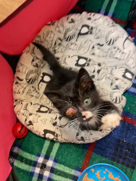


Disclaimer: kitten has already seen a vet, enucleation is planned as soon as she hits their minimum weight. Just asking as a vet student who feels like they have way too many little bits of info floating around in their brain to be sure about anything lol.
Kitten was dropped off at shelter with 0 history (of course), and my knowledge on this case is a little vague. Is this likely just from an infection left too long, that probably sealed the eye shut at some point and worsened? (Like, viral -> bacterial?) It looks it’s proptosed to me, but I’m also not sure if that’s just massive swelling. If it IS proptosed, is that a common sequelae of severe infection? Or do you really only get that with trauma in cats, and then secondary infection?
Not sure if my thinking is way off base, just looking for some general insight into how these eyes end up looking so gnarly. I feel like we’ve had quite a few dropped off this year. Thank you!
Hi, it's Sueanoi here.
I'm glad the kitten is getting some vet attention now. I hope the healing goes as smooth as it can.
As to answer your question, I don't think that's proptosis (eyeball goes out of orbit). I think it is ruptured cornea that has severe inflammation on top.
Anything at all can cause a secondary corneal injury if the eye is irritated, causing the animal to scratch it and worsening the lesion. Over time, superficial ulcers can become perforated.
Over here in SEA, young stray cats are very often infected with feline herpes. It is a very common cause of blindness (as the last consequence of virus-induced ulcer) here.
Your thinking isn't entirely off base, because virus -> bacteria -> catastrophic consequense IS probably correct.
Once it is time to enucleate, I suggest you observe the surgery. I am quite certain that the globe itself is still sitting within the orbit. The things that are coming out of the cat's head is the eyeball's insides that are spilled out of the ruptured cornea.
Keep clean and control infection. There is a chance that the cat won't even need the surgery if the eye shrink down and become enophalmos before it reaches minumum weight (which might take weeks...whichever happen first). Downside of keeping the shrunk eye is having to keep it clean for the rest of its life. So if that is a factor for the cat getting an adoption, removing it anyway for ease of care is still a valid choice.
Best of luck!
45 notes
·
View notes
Text
Being rendered helpless (PANOPTICON)
• Rita Ora's thumb (Encounter for aftercare following multiple organ transplant)
• Florence Welch's thumb (Laceration with foreign body of right ring finger with damage to nail)
• Winona Ryder's thumb (Secondary lacrimal gland atrophy)
• Lucy Hale's thumb (Failure in dosage during unspecified surgical and medical care)
• Conan O'Brien's thumb (Influenza due to other identified influenza virus with otitis media)
• Tyra Banks's thumb (Malignant neoplasm of overlapping sites of other and unspecified parts of mouth)
• AnnaSophia Robb's thumb (Laceration of extensor muscle, fascia and tendon of left middle finger at forearm level)
• Minka Kelly's thumb (Acute tonsillitis, unspecified)
• Djimon Hounsou's thumb (Cyst and mucocele of nose and nasal sinus)
• Forest Whitaker's thumb (Meningococcal myocarditis)
• Jimmy Buffett's thumb (Other disorders of continuity of bone, right radius)
• Kate Bosworth's thumb (Other hyperparathyroidism)
• Kristen Bell's thumb (Solitary bone cyst, left ulna and radius)
• Matt Bomer's thumb (Laceration of other muscles, fascia and tendons at shoulder and upper arm level, unspecified arm)
• Prince Harry's thumb (Laceration without foreign body of back wall of thorax without penetration into thoracic cavity)
• Avril Lavigne's thumb (Calcification and ossification of muscle)
• Demi Lovato's thumb (Nondisplaced fracture of lateral condyle of unspecified femur)
• Carmen Electra's thumb (Salter Harris Type III physeal fracture of upper end of humerus, left arm)
• Mary-Louise Parker's thumb (Atherosclerosis of other type of bypass graft(s) of the extremities with intermittent claudication, left leg)
• Vince Vaughn's thumb (Toxic effect of contact with other venomous marine animals, assault)
• Sean Lennon's thumb (Unspecified open wound of left front wall of thorax without penetration into thoracic cavity)
• Tate Donovan's thumb (Osseous and subluxation stenosis of intervertebral foramina of abdomen and other regions)
• Jennifer Aniston's thumb (Alcohol abuse with intoxication)
• Zachary Quinto's thumb (Mooren's corneal ulcer, unspecified eye)
• Tracy Morgan's thumb (Preterm labor without delivery, unspecified trimester)
• Jenna Elfman's thumb (Inflammatory polyneuropathy, unspecified)
• Kaley Cuoco-Sweeting's thumb (Perforated corneal ulcer, unspecified eye)
• DJ AM's thumb (Kaschin-Beck disease, left knee)
• Gordon Ramsay's thumb (Unspecified injury of extensor muscle, fascia and tendon of right index finger at forearm level)
• Elle Fanning's thumb (Benign neoplasm of connective and other soft tissue of unspecified upper limb, including shoulder)
• Scott Speedman's thumb (Encounter for routine postpartum follow-up)
• Curtis Stone's thumb (Swimmer's ear, left ear)
• Uma Thurman's thumb (Altered mental status, unspecified)
• Khloe Kardashian's thumb (Retinal hemorrhage, left eye)
• Maria Menounos's thumb (Passenger in three-wheeled motor vehicle injured in collision with fixed or stationary object in nontraffic accident)
• Miranda Kerr's thumb (Other combined immunodeficiencies)
• Brooklyn Decker's thumb (Atherosclerosis of other type of bypass graft(s) of the extremities with intermittent claudication, left leg)
• Ellie Goulding's thumb (Osteonecrosis in diseases classified elsewhere, thigh)
• Bethenny Frankel's thumb (Other chronic hematogenous osteomyelitis, left humerus)
• Judi Dench's thumb (Resistance to unspecified beta lactam antibiotics)
2 notes
·
View notes
Text
Can Lasik Surgery Correct Vision Problems Caused by Corneal Melt?
Lasik surgery is a popular procedure for correcting vision problems, particularly for those with refractive errors. But can it address more complex issues like corneal melt? This is a common question, especially for patients dealing with corneal conditions. In this article, I, Dr. Gitansha Shreyas Sachdev, will explain how Lasik surgery works and whether it can help in cases of corneal melt. If you're seeking Lasik Surgery in Bangalore, we offer comprehensive solutions at The Eye Foundation.
Understanding Corneal MeltCorneal melt is a rare condition where the cornea—the clear outer layer of the eye—starts to thin and degrade. This can lead to severe vision problems and discomfort. Causes include autoimmune disorders, infections, and, in some cases, eye surgeries. The key issue with corneal melt is that it affects the structural integrity of the cornea, which is crucial for focusing light onto the retina.
What is Lasik Surgery?Lasik, or Laser-Assisted in Situ Keratomileusis, is a type of eye surgery that reshapes the cornea to correct vision problems such as myopia (nearsightedness), hyperopia (farsightedness), and astigmatism. During the procedure, a laser is used to remove a precise amount of corneal tissue, allowing the light to focus correctly on the retina, thereby improving vision.
Can Lasik Address Vision Problems from Corneal Melt?Lasik surgery is highly effective for refractive errors, but it may not be suitable for patients with corneal melt. The structural damage caused by corneal melt could complicate the reshaping process, as the cornea may be too thin or weakened. Attempting Lasik on an already compromised cornea can lead to further complications, such as worsening vision or even corneal perforation.
Who is Eligible for Lasik Surgery?While Lasik surgery may not be the right choice for those with corneal melt, it remains an excellent option for individuals with refractive errors. To determine if you're a good candidate for Lasik, we consider several factors:
Age: Most patients should be 18 or older.
Stable Vision: Your prescription should be stable for at least one year.
Healthy Eyes: You should be free from conditions like dry eye syndrome, glaucoma, or cataracts.
Step-by-Step Lasik Surgery ProcessIf you're a suitable candidate for Lasik, here’s what you can expect from the procedure:
Initial Consultation: We perform a comprehensive eye exam to determine your eligibility.
Creating the Flap: A thin flap is created in the cornea using either a microkeratome or a femtosecond laser.
Reshaping the Cornea: The laser reshapes the underlying corneal tissue, correcting the refractive error.
Closing the Flap: The flap is carefully placed back without the need for stitches.
Post-Operative Care: You'll need to follow a recovery regimen, including the use of prescribed eye drops.
Post-Surgery Care and RecoveryAfter Lasik surgery, most patients experience improved vision within 24 hours. However, it’s essential to follow post-surgery guidelines to ensure a smooth recovery:
Avoid rubbing your eyes.
Use prescribed eye drops to prevent infection.
Attend follow-up appointments for monitoring.
Key TakeawaysLasik surgery is a powerful tool for correcting refractive vision issues, but it is not a solution for corneal melt. Patients with corneal melt should explore other treatment options, such as corneal collagen cross-linking or a corneal transplant. If you're considering Lasik Surgery in Bangalore, our team at The Eye Foundation can provide expert guidance and personalized treatment plans.
#lasik surgery bangalore#lasik surgeon#lasik eye surgery#lasik treatment#lasik surgery#lasik treatment bangalore
0 notes
Text
Rising Adoption To Treat Ophthalmic Disorders Projected to boost growth of the Amniotic Membrane Market
The global Amniotic Membrane Market is estimated to be valued at US$ 3.62 Bn in 2023 and is expected to exhibit a CAGR of 13% over the forecast period 2023 to 2030, as highlighted in a new report published by Coherent Market Insights.
Market Overview:
The amniotic membrane is the innermost layer of the placenta which consists of epithelial cells, a thick basement membrane, and an avascular stroma. It possesses properties such as anti-inflammatory, anti-scarring, antimicrobial, anti-angiogenic and pain-relieving properties. This makes it ideal for use in wound healing applications including wounds, burns, ophthalmic disorders, surgical wounds etc. It acts as a biological scaffold and releases growth factors that aid in tissue regeneration. Compared to synthetic skin substitutes or skin grafts, amniotic membrane grafts have lesser probability of developing infection and rejection. With increasing cases of trauma, accidents and chronic wounds worldwide, the demand for effective wound care treatments is growing steadily. This is expected to propel the amniotic membrane market growth over the forecast period.
Market key trends:
One of the key trends gaining traction in the amniotic membrane market is the rising adoption of amniotic membrane for ophthalmic disorders. Amniotic membrane plays a crucial role in ocular surface reconstruction due to its anti-angiogenic, anti-inflammatory and anti-scarring properties. It is increasingly being used to treat ocular disorders like keratitis, corneal ulcers, persistent epithelial defects and corneal perforations. Also, continuous efforts by market players to develop new amniotic membrane products through advanced processing techniques like cryopreservation, lyophilization etc. is expected to drive the market growth. Development of amniotic membrane tissue engineered products for regenerative therapies is another emerging trend in the market.
Porter’s Analysis
Threat of new entrants: Low, as the market requires high capital investment and regulatory approvals for entering.
Bargaining power of buyers: Moderate, as the presence of substitute products provides options to buyers.
Bargaining power of suppliers: High, as raw materials have limited sources of supply globally.
Threat of new substitutes: Moderate, as new substitutes are emerging but amniotic membrane has established use cases over years.
Competitive rivalry: High, as the key players compete on the basis of quality, price and technology.
Key Takeaways
The global amniotic membrane market size is expected to witness high growth at a CAGR of 13% over the forecast period 2023 to 2030, due to increasing cases of ocular surface disorders.
Regional analysis: North America dominates the global market currently owing to increasing healthcare expenditure and growing patients' population. However, Asia Pacific is expected to grow at the fastest pace during the forecast period with rising medical tourism and improving healthcare infrastructure in emerging countries.
Key players operating in the amniotic membrane market include Smith & Nephew, Applied Biologics, Organogenesis Holdings Inc., Osiris Therapeutics, Inc., Alliqua BioMedical, Inc., Amnio Technology, LLC, Next Biosciences, Integra LifeSciences, MiMedx, LifeCell International, Human Regenerative Technologies, LLC, Amniox Medical, Inc, Skye Biologics Inc., Wright Medical, TissueTech, Katena Products, Inc., NuVision Biotherapies, Surgenex, and Ventris Medical. Key players are focused on new product launches and regulatory approvals to strengthen their market position.
0 notes
Text
Eyebrow Lamination
Eyebrow Lamination is a semi-permanent treatment that’s a bit like a hair perm for your eyebrows. It’s best left to the professionals as many at-home products are too harsh, leaving your brows dry and brittle.
Typically, it lasts 4-6 weeks but can last up to 8 with proper care. It can also help you reshape and change the direction of your natural brow hairs, creating a more flattering arch.
What is it?
Eyebrow lamination is a non-invasive treatment that involves styling your brow hairs to look fuller and neater. It’s often paired with a brow tint, which can add color and make the brows appear more bushy. The procedure can be used to fill in sparse or thinning brows, and it’s also great for people who have lost their natural eyebrow shape due to over-tweezing, waxing or baldness.
Gafni warns against trying brow lamination at home, as it could cause damage to the delicate eyebrow skin if the chemical is accidentally applied in the wrong place. She says that if the brow lamination solution drips into your eyes, it may cause chemical burns or corneal perforation, both of which can be permanent. Instead, she suggests booking the treatment at a professional salon that specialises in it.
How does it work?
Eyebrow lamination is a two-step treatment that works well on coarse or unruly eyebrows, as well as thin ones that have been affected by overplucking or hair loss. The first step involves a cream that breaks down the bonds in the individual brow hairs, which relaxes and straightens them. From there, the stylist can reshape the brows into the desired appearance confirmed in the consultation.
The next step involves another product that rebuilds the brow hairs' broken bonds, setting them into their new shape. As a result, the brows appear thicker and fuller. This makes it a great option for those who want to ditch fillers and go more natural. The results will last for as long as the natural brow hair growth cycle, and can be maintained with products like a spoolie brush or a clear brow gel.
How long does it last?
A few weeks after the treatment, you’ll notice that your brows will start to relax a little. This is because hairs naturally fall out and grow back with their own natural growth cycle. The good news is that you can extend your lamination results with dedicated aftercare, like using a hydrating oil to help coach the hairs to grow in the desired direction.
Eyebrow lamination is a great option for those who want thicker, fuller brows or to camouflage thinning, patchiness or sparse areas of their brows. It also offers a more precise and neat appearance than a regular brow tint.
However, it is not recommended to perform this service during pregnancy and breastfeeding. This is because hormones can impact the outcome of the treatment.
Can I remove it?
Eyebrow lamination gives brows a beautiful brushed-up appearance, and it saves you grooming time in the morning. It is a semi-permanent treatment that lasts for 6 weeks. It is a great option for those with unruly, easily-tangled or curly eyebrows. It also helps make thin brows look fuller and thicker.
However, many people complain about their brows getting frizzy after the procedure. This is usually due to the type of perm solution used. Thioglycolic acid is the most common type of perm solution. This chemical causes the brow hairs to be raised to a very high pH and then neutralized.
The best way to avoid your brows becoming frizzy is to use a nourishing oil. Marris recommends using her Brow Code Brow Gold Nourishing Growth Oil, which can be purchased online.
What are the risks?
Remember soap brows, the semi-permanent eyebrow trend that was all over TikTok for a while? Well, if you’re thinking of giving this trend a go with your beautician, make sure to ask about their experience with lamination. The formulas used in this process can be harsh and dry out brow hairs, leading to breakage and weaker strands. They also contain chemicals with questionable safety records and can irritate the skin.
If you have a sensitivity to these ingredients, it’s not recommended to get lamination done. And don’t forget to do a patch test before the procedure to check for any allergic reactions. A more serious risk is eye damage if the chemicals run into your eyes. This is why it’s important to choose a salon that has the proper safety procedures in place.
0 notes
Text
Okay so what I had had dismissed as annoying eye allergies turned out to be a bigger deal after all. Like almost rushed into emergency surgery bigger. Thankfully I was offered and tried a much less invasive and nerve wracking option that seems to be doing the trick. Basically I had a corneal ulcer that got perforated and they "glued" it back together. Now just have to see how it heals up and hope for minimal scarring.

I don't normally look this cool, got the doctor ordered perma-shades to blame 😎😋
133 notes
·
View notes
Text
Beckoning cat without left hand: Life history of Choma-1(1/2)

This painting is a portrait of Choma that I unknowingly drew about two months before Choma's death.
It is an oil painting with a title such as "Handless Maneki Neko".
In 1993, I was surprised when my father, who had a ruthless attitude toward living things, said that he would keep a "cat". I thought, "A man who is as cold as my father keep a cat?"
The cat I saw for the first time was a really small cat, and I thought, "What is this little one?"
I noticed and received one of the kittens born by the clerk of the company where my father was the factory manager. And this cat is a cat that has "Scottish Fold" in one parent, and the place where the ears originally hang down, but the characteristic did not appear in the cat himself.
My father gave the name "Choma". It seems that it means eating food little by little. That's why my father also called it "Chomako" and "Guitsukotan".
They are together when they go to bed.
And Choma grew up, given cat food and occasionally fish sashimi.
In the case of Choma, the food that should be served at the set time ,but served he was screaming, so he grew bigger and became a heavyweight cat. Approximately 9 kg.
And, as a male cat’s nature, he went on an expedition to the outside of our house, cultivated territory, and returned triumphantly. Such a choma has a rival, and he comes home after a quarrel with a lot of lives, so the injury was usual. For example, "corneal perforation" is a fear of blindness, so I took him to a veterinary clinic and received eye drops. Also, in the case of cats, the surface of the injured part will soon heal and lesions will remain in the body, so it was necessary to pay attention to this as well.
Also, at my home, We was feeding the stray cats as well, so Choma was kicking them down as if he was looking down on them. It was an arrogant attitude. Once, Choma was hitting a poor cat "Knock" who escaped to the outdoor unit of the air conditioner, so I once held a Choma and let the knock escape in the meantime, saying "You also like bullying the weak". ..
Among these stray cats, there was a gentle brother-like cat, who lived while caring with his colleagues. We used to call it "Know". This cat's brother has a light coat color, but the coat color is dark, so he called it "Know (dark)". The other is "tan (light)". Easy nomenclature.
January 10, 1996 came to my house doing so.
On January 10, 1996, something happened to Choma. In the daytime, I heard some kind of squeaky voice, so there was a Choma in the doorway room ... But if I look closely, the wrist of his left hand is "twisted", and it looks horrible.
I had to take him to a veterinary clinic right away, but I was surprised ,when he ate food for such a time .
I heard at a veterinary clinic that he can't make an "artificial limb", and if the front leg remains halfway, the bones will be exposed, so we have to take the left front leg together.
Choma's surgery was performed there, and Choma became three legs.
After the accident, the appearance of "Know" disappeared, so I suspect that Choma was bullying Know, but he tampered with the artificial rotating equipment and screwed it. "Know" would have robbed one hand of Choma in exchange for his life.
To be continued...
#twisted#choma#Beckoning cat#Scottish Fold#male cat’s nature#territory#the injury was usual#stray cats#squeaky voice#veterinary clinic#the wrist of his left hand is#Maneki Neko#rei morishita
22 notes
·
View notes
Text
So 2 nights ago I was on call and was in until midnight for a horse with a melting corneal ulcer. Owners opted for medical management despite a huge ulcer and worse prognosis due to fungal infection.
After this morning, I now know what it looks like to see the iris prolapse as an eye perforates.
Also what the vitreus humor looks like shooting out when a standing equine enucleation has its draft horse head yanked up out of reach.
It's been a wild Friday.
#veterinary student#vet med#vetblr#vet school#future dvm#ophtho#ophthalmology#7/8#on call#ulcer#equine#horses are fools
14 notes
·
View notes
Text
Efficacy of Amniotic Membrane Transplantation in Refractory Infective Keratitis Leading to Stromal Thinning, Descematocele and Perforations- Juniper Publishers
Juniper Publishers- JOJ Ophthalmology
Introduction
Diseases affecting the cornea are a major cause of blindness all over the world, second only to cataract in overall importance [1]. One of the commonest corneal causes is Infectious Keratitis. The prevalence of blindness directly resulting from complications of Infective Keratitis is estimated to be 5% [2]. Cases refractory to the medical therapy requires urgent surgical intervention to retrieve the vision and most importantly to salvage eye. Available surgical management in refractory keratitis cases include tissue adhesives, Bandage Contact Lenses (BCL), penetrating or lamellar keratoplasty [3] patch grafts, or conjunctival flaps. Unfortunately, these therapies are associated with a considerable number of complications and address only the tectonic problem, without solving the ongoing infection and inflammation. BCL and conjunctival flaps being a temporary measure does not provide with new collagen to improve corneal thickness and stabilize the cornea. For such situations Penetrating Keratoplasty (PK), Lamellar Keratoplasty (LK) or patch grafts was the only option and is still being used widely. PK and patch grafts performed to seal a corneal perforation may be complicated with synechiae, glaucoma, uveitis, and graft failure in the setting of an inflamed or infected eye [4]. Recurrence of infection in corneal grafts is also challenged. LK being difficult to perform may result in a double chamber between the donor and recipient cornea in some cases. Tissue adhesives may dislodge and are used as a temporary measure, obviating the need for a PK within a few days [5,6].
Preserved human amnion has been successfully used as a biological bandage, promoter of epithelialization, inhibitor of inflammation and angiogenesis, as well as a carrier for ex vivo cultured limbal stem cells [7]. Amniotic Membrane Transplant (AMT) offers the advantage of avoiding potential allograft rejection. Even if corneal transplantation is needed, the success rate is improved if performed on an eye that underwent AMT reducing inflammation [8,9]. Amniotic Membrane (AM) integrates in cornea and thus can be used as a treatment for corneal perforation by restoring corneal stromal thickness so that emergency PK can be avoided, as suitable donor corneal button availability is difficult in every place. Therefore, an alternative management for various stages of infectious keratitis including deep refractory stromal ulcers, descematocele and corneal perforations is reconstruction of the surface with AMT adjuvant with appropriate antimicrobials and supportive medications. In this prospective study AMT in various gravities refractory infective keratitis has been attempted to understand the efficiencies and limitations associated with it.
Methods
A prospective, interventional study was done on 150 eyes of 150 patients. All patients with refractory (unresponsive to conventional treatments significantly for more than 2 weeks) infective keratitis, advanced infectious keratitis with descematocele and corneal perforation requiring urgent concealment to salvage the eye, were treated with single or multi layered AMT. Patients with non-infective ulcers and perforations were excluded from the study. Corneal ulcer was graded 1-5 according to the depth of corneal involvement on slit lamp biomicroscopy (Table 1). Microbial investigations (staining for bacteria and fungus with culture-sensitivity) were done and antimicrobials started accordingly. B-scan ultra sonography was done in hazy media to rule out involvement of posterior segment. Any systemic (diabetes) or ocular (dacryocystitis) conditions hindering the healing of ulcer or triggering the infection were investigated and managed.
On basis of slit lamp examination at the site of most impact.
Technique
Surgery was performed preferably under sub conjunctival or peribulbar anesthesia. In children or uncooperative patients general anesthesia was used. Debridement of the necrotic tissue was done from and around the ulcer bed. Care was taken to remove the pseudo cornea over the perforation at the end of debridement to prevent leaking of aqueous and thus allowing proper keratectomy. Single layer preserved AM was used in cases of deep stromal ulcer. AM with epithelial side up was spread over the ulcer and trimmed to fit the ulcer. It was secured with continuous or interrupted 10-0 monofilament nylon suture. Descematocele and small corneal perforations up to 4mm were treated with multilayer AMT owing to deep corneal involvement. A sheet of AM, folded over it-self with epithelial side out, filled the ulcer crater and anchored to the healthy ulcer margin with interrupted 10-0 nylon suture. It was covered with a single sheet of AM similarly as in cases of deep stromal ulcers. In large corneal perforations of 4-6mm with extensive surrounding stromal necrosis, margins were not sturdy to hold the suture and there was a risk of cutting-off a corneal bite. In such cases single layer was sutured at limbus to at least provide tectonic support to the eyeball and delaying the need for PK. Side port or paracentasis was made in cases hypopyon and corneal perforation to reform the anterior chamber with air and reposit the prolapsed iris with help of spatula. Anterior synechiae if present were broken to prevent formation of adherent leucoma and thereby secondary glaucoma. Hypopyon if present was washed through the side port and intracameral antibiotic or antifungal was also injected according to sensitivity. At the end a BCL was placed over the cornea to prevent irritation from corneal sutures and maintaining AM in place. Antimicrobial, cycloplegics, ocular hypotensive and lubricating drops were continued along with systemic supportive therapy. Frequent follow-ups were done weekly for 1 month, biweekly till 3 months and monthly till 6 months. Efficacy was monitored on basis of improvement in symptoms and visual acuity, healing of the ulcer by re-epithelization and formation of anterior chamber, achievement of corneal transparency and corneal thickness. Accordingly patient's outcome was described as satisfactory, intermediate and failure (Table 2).
Observation and Results
Keratitis was classified (Table 1) according to the depth of the cornea involved into 5 grades. Grades 1 and 2 responded well with medical management, therefore did not require AMT. Grades 3-5 with deeper corneal penetration of infection did not heal merely with medical management, there was an apprehension of corneal thinning and progression of infection, which required AMT. Of the 150 patients who underwent AMT, 55 (36.67%) were deep stromal ulcers, 25 (16.66%) were descematocele and maximum 70 (46.67%) patients were of corneal perforation ranging from 1-6mm. There was no age group or gender preponderance. Symptoms of redness, pain, watering and foreign body sensation (FBS) were collectively present in all the cases. Lid oedema and photophobia were also present in majority of the cases (70.6% and 90% respectively).Presence of discharge was seen in moderately less cases (30%). ranging between 1-2mm and 10 cases (20%) had hypopyon of Hypopyon was present in total 50 (33.3 %) cases where 10 cases >2mm (Table 3).
Single layer AMT was done in total 85 cases, all 55 cases of deep stromal ulcer and 30 cases of corneal perforation >4mm with extensive necrosis to provide tectonic support to maintain integrity to eyeball. Roofing with multilayer technique was done in 65 cases, all 25 cases of descematocele and 40 cases of corneal perforation >4mm in largest dimension where neighboring corneal tissue was healthy to hold the corneal sutures (Table 3). Patients were observed in repeated postoperative days. Rapid descent of symptoms was observed after the AMT. There was drastic improvement in pain, lid oedema, FBS and discharge in the first week. Symptoms were barely present in few cases by 1 month, which totally recovered by 3 months in all the cases (Figure 1).
Corneal transparency graded from 0 (leucomatous opacity) to +4 (clear cornea, with no haze) was measured objectively at the site of most impact on slit lamp (Table 4). Improvement was seen in 105 of 150 cases and was statistically significant (p=0.016). However none of the cases improved to +4 transparency that is totally clear cornea (Table 5). Visual acuity was recorded before and after 6 months of treatment in 145 of 150 cases as 5 cases of fungal ulcer failed to heal with AMT (Table 6). Improvement in BCVA when taken collaborate, was extremely significant (p >0.0001). Mild to moderate complications were faced during the entire course of treatment. They were shallow anterior chamber in 5 cases in perforation which was tackled with air injection in anterior chamber and breaking anterior synechiae. Hemorrhage beneath AM in five cases which resolved spontaneously. Graft retraction was seen in five cases for which repeat AMT was done. Hypopyon developed in 10 cases and did not resolve with topical therapy was managed with anterior chamber wash and intracameral moxifloxacin and amphotericin-B respectively (Table 7). Hypopyon did not redevelop in these cases. All the complications were successfully managed with appropriate treatment with no recurrence and good results. Also no re-infection was noted. Graft melting and corneal perforation was seen in 5 cases of fulminant fungal ulcer and required urgent therapeutic PK.
Satisfactory results were seen in 100 of 150 eyes (66.67%), intermediate results seen in 45 cases (23.33%). Failure was noted in 5 cases (3.33%) of fulminant fungal ulcers that showed subsequent corneal perforation requiring Therapeutic PK (Table 8). All the cases in intermediate category which also required subsequent intervention, healed with stable cornea. Thus, successful results were seen in 145 of 150 cases (96.67%) of which in 30 cases subsequent penetrating keratoplasty was done for leucomatous corneal opacity obscuring the visual axis left after healed ulcer (Figure 2).
Discussion
Approximately one-third of cases of infective keratitis require surgical interventions at the acute stage to prevent perforation or spreading of infection [10-14]. Keratoplasty being majorly followed in such situation faces a limitation of availability of good quality donor corneas, mainly in developing countries, recurrence of infection, difficulty in technique and graft rejection. Moreover, for fungal keratitis PK is technique dependent and may also carry a risk of recurrent infection [15].
Thus AMT is sought as an alternative, which has been extensively reported in ophthalmology literature [16-19]. AMT offers the advantage of stimulating re-epithelization, preventing neovascularization and scar formation and avoiding potential allograft rejection. Even if corneal transplantation is needed, the success rate is improved if performed on an eye with reduce infection and inflammation, this can be achieved with AMT [8,9]. In present study complete epithelization was noted in 145 of 150 cases, that is 96.67% success rate. Similar to our study, Chen et al. [20], showed 82.61% success rate, 4 of 23 cases in there study faced AM melting and graft failure requiring therapeutic PK in 3 and delayed healing with vascularization in the other. Kim et al. [21] used multilayer AM in cases of descematocele and corneal perforation. Corneal surface was healed successfully in all cases, and no recurrence of infection or rejection was experienced. Hanada et al. [22] used multiple layers of AM for deeper stromal ulcers down to descemetocele, to restore the normal corneal thickness as well as in corneal perforations from 0.5 to 3mm with or without additional tissue adhesive with high success rates (73-93%). In present study corneal perforations in cases of infectious keratitis up to 6mm have been treated successfully with AMT alone, and 100% corneal epithelization with more than 50% corneal thickness have been achieved in all 70 cases of perforation. In a series by Heiligenhaus et al. [23]. Seven patients with herpes simplex virus or varicella zoster- induced severe ulcerative keratitis, 5 of 7 eyes healed after first AMT [23]. In another study, stromal defect was filled up with multilayer technique proved to be better than monolayer procedure [22,24,25].
In present study 70% showed significant improvement in corneal transparency and increasing corneal transparency improves the best-corrected visual acuity further emphasizing the healing properties of AMT. Chen et al. [20], preserved useful vision after AMT in cases of fungal keratitis in 52.2% eyes. Kim et al. [21], reported 21 cases of successful AMT in infectious keratitis, in which visual acuity increased except for 5 cases because of irreversible corneal opacity. AMT has come up as a very effective managing technique for refractory ulcers. It aids in permanent healing of the refractory infective keratitis and prepares the cornea for definitive reconstructive procedure if required (Figure 3).
Conclusion
We have found that AMT represents a viable method of treatment to promote healing and prevent progressive melting of refractory infectious keratitis. Besides being cost-effective it’s easy to perform, with a short learning curve. Thus, it might be considered a first-line surgical technique when maximal medical treatment has failed.
For more Open Access Journals in Juniper Publishers please click on: https://juniperpublishers.com
For more articles in JOJ Ophthalmology (JOJO) please click on: https://juniperpublishers.com/jojo/index.php
For more Open Access Journals please click on: https://juniperpublishers.com
0 notes
Text
April 4 Cases
1. 406 with teary and itchy eyes, told to use HC BID and Flarex. No microwave so I suggested a baked potato 😂
2. Infiltrate in AOK patient!! Epi defect! Used Vigamox QID. Could not bill 402 because was due for a 404.
Should I have increased DOSAGE of Vigamox to Q2H because of MK risk?
3. 404 with two lower lid styes x months! Had an uncle who had recurrent styes and was put on antibiotics long-term. Used doxycycline 100mg x2wk, counselled ADR of nausea.
4. BCL case - probability of CL EW infection (10-20/10 000 in EW SCL) vs probability of corneal perforation from entropion? Counselled no tap water or swimming in CLs. DO I ADD PROPHYLACTIC ANTIBIOTIC OVER EW CL?
0 notes
Text
Confess and be hanged
Kathy Griffin's elbow (Other congenital malformations of hair)
Dave Navarro's forehead (Subluxation of lens, unspecified eye)
Jessica Biel's eye (Other hammer toe(s) (acquired), left foot)
James Franco's fist (Solitary bone cyst, left ulna and radius)
Simon Doonan's thigh (Malignant neoplasm of left orbit)
Carson Palmer's head (School (private) (public) (state) as the place of occurrence of the external cause)
Pitbull's eye (Chondrolysis, hip)
Kevin Federline's eye (Osseous and subluxation stenosis of intervertebral foramina of abdomen and other regions)
Tate Donovan's thigh (Chronic myeloid leukemia, BCR/ABL-positive, in remission)
Ryan Gosling's arm (Pedal cycle passenger injured in collision with fixed or stationary object in traffic accident)
Sean Combs's neck (Mixed pediculosis and phthiriasis)
Katharine McPhee's chin (Calcific tendinitis, right lower leg)
Katrina Bowden's back (Kernicterus, unspecified)
Balthazar Getty's hair (Toxic effect of contact with other venomous marine animals, assault)
Elizabeth Taylor's ear (Displaced trimalleolar fracture of left lower leg)
Kelsey Grammer's eye (Major laceration of left kidney)
Kerry Diamond's neck (Scrotal transposition)
Jason Lee's wrist (Papyraceous fetus, first trimester)
Josh Holloway's upper arm (Activity, swimming)
Desiree Hartsock's ear (Swimmer's ear, left ear)
Jared Leto's eyebrow (Pathological fracture, right hand)
Rumer Willis's eye (Lesion of plantar nerve)
Ramona Singer's arm (Other specified injury of intrinsic muscle and tendon at ankle and foot level, left foot)
Emily VanCamp's calf (Nicotine dependence, cigarettes, with withdrawal)
Jane Krakowski's fist (Other unilateral secondary osteoarthritis of hip)
Vince Vaughn's lower leg (Unspecified complication following infusion and therapeutic injection)
Olivia Palermo's shoulder (Laceration without foreign body of right back wall of thorax with penetration into thoracic cavity)
Russell Brand's wrist (Malignant neoplasm of left orbit)
Jackson Rathbone's belly (Primary cyst of pars plana, unspecified eye)
Garth Brooks's eyebrow (Nondisplaced fracture of anterior process of left calcaneus)
Adrian Grenier's nose (Military operations involving flamethrower, civilian)
Jesse Tyler Ferguson's hair (Retinal hemorrhage, left eye)
Martin Lawrence's ankle (Hemorrhagic disease of newborn)
Spencer Pratt's neck (Perforated corneal ulcer, unspecified eye)
Ashley Hebert's bottom (Major laceration of left kidney)
Hugh Jackman's bottom (Laceration of radial artery at wrist and hand level of left arm)
Paris Hilton's chin (Preterm labor without delivery, unspecified trimester)
Simon Cowell's arm (Contusion of small intestine)
Tila Tequila's cheek (Other superficial bite of hand of unspecified hand)
Jennifer Grey's toe (Injury of quadriceps muscle, fascia and tendon)
Brody Jenner's hip (Laceration without foreign body of back wall of thorax without penetration into thoracic cavity)
Ciara's hair (Diffuse cystic mastopathy of unspecified breast)
Molly Sims's chin (Urticaria due to cold and heat)
Luke Bryan's buttocks (Urticaria due to cold and heat)
Richard Gere's breast (Endometriosis of pelvic peritoneum)
Jensen Ackles's calf (Other ulcerative colitis with intestinal obstruction)
Teresa Giudice's head (Laceration of extensor muscle, fascia and tendon of left middle finger at forearm level)
Stavros Niarchos III's ear (Striatonigral degeneration)
Winona Ryder's thumb (Acute embolism and thrombosis of right femoral vein)
Scott Disick's forearm (Extranodal NK/T-cell lymphoma, nasal type)
3 notes
·
View notes
Text
Juniper Publishers- JOJ Ophthalmology Nasolacrimal Duct Obstruction Review-JOJ Ophthalmology
Introduction
The lacrimal system comprises two components the main and accessory lacrimal glands and their secretions and the lacrimal excretory system [1]. The lacrimal excretory system is divided into the proximal and distal sections. The proximal section includes the punctum, canaliculus, and the common canaliculus [2,3]. The distal lacrimal drainage system consists of the lacrimal sac and the nasolacrimal duct that finally ends under the inferior turbinate and empties into the inferior meatus [2].
The precorneal tear film is composed of aqueous, mucinous and oily components and is necessary for the maintenance of the cornea as well as the maintenance of the ocular surface epithelium. More than 90% of the lacrimal fluid is removed by the excretory system, whereas less than 10% evaporates between blinks. Outflow is mainly regulated by the pumping effect of the orbicularis oculi muscle (Horner muscle) [3,4]. Tears are thus drawn into the lacrimal excretory system after each blink. The passage of tears down the nasolacrimal duct is influenced by gravity, evaporation in the nose, and inspiration and expiration.
Diagnosis
A detailed history of any systemic or topical medication, surgery, trauma or scarring, and infection must be obtained. It is valuable to grade the severity of epiphora using a uniform grading system such as the Munk scale [5]. Slit lamp examination starts with recognizing the papilla, presence of a membrane or fibrosis over the punctum, punctum size, tear meniscus height, eyelid margin, conjunctiva around the punctum, eyelid malposition, position of the punctum in the tear lake, and any sign of previous surgery. The Schirmer test [6], tear break up time [7], ocular surface staining, and tear meniscus height will rule out any associated ocular surface abnormalities. Abnormal dye disappearance test is a very maneuver to assess abnormal tear drainage system and is especially helpful in pediatric patients [7].
Congenital Nasolacrimal Duct Obstruction
Congenital nasolacrimal duct obstruction is the first cause of pediatric epiphora. Other causes include congenital punctum and canaliculus stenosis and/or atresia, nasal malformations and craneofacial abnormalities. It is frequently seen at birth due to lack of perforation of the valve of Hasner or an inferior and distal nasolacrimal duct opening failure. At birth, half of the nasolacrimal new born pathways are not permeable. A spontaneous apoptosis mechanism takes place between 3rd- 4th weeks after birth. Nevertheless, the obstruction persists in approximately 20% of the patients. Symptoms of congenital nasolacrimal duct obstruction consist of epiphora and dacryocystitis. The diagnosis is easily made in the office by observing epiphora and mattering of the eyelashes (Figure 1). It can be confirmed by compression over the nasolacrimal sac, which results in regurgitation of mucopurulent material in those patients who have developed chronic dacryocystitis. Instillation of 2% fluorescein dye and observation of abnormally delayed passage from de cul-de-sac is helpful in confirming the diagnosis.This is a rare entity presented at birth or within the fourth week after birth. The blockage Rosenmüller valve. An edematous, tender and red mass below the medial canthal tendon will be clinically found (Figure 2). Conservative management (medical treatment). If dacryocele is initially sterile, all patients must be treated with warm compresses, local massage and topical antibiotics [8]. Those infected will be treated with broad spectrum intravenous antibiotic therapy [9]. Local massage by pushing down the lacrimal sac is useful and accelerates the lumen duct perforation process (Figure 3). Controversy exists whether conservative management or early probing for decompression are preferable. Surgical Management of Congenital Nasolacrimal ObstructionProbing consist by introducing a thin metal probe into the lacrimal punctum trough the nasolacrimal pathway, producing a mechanical opening in the obstruction site. It is an operating room procedure under general anesthesia. The ideal time is controversial, most of the time the procedure is performed around the first year of life for those patients that did not show spontaneous improvement or despite conservative treatment [10].Close dacryointubation is performed by placement of silicone stents through the superior and inferior canaliculus and down to the nasolacrimal duct. This dilates the inferior meatus. The duration of the stent employment ranges from 6 weeks to 6 months [10]. Dacryocystorhinostomy surgical procedure involves the removal of bone adjacent to the lacrimal sac draining directly into the nasal cavity and it is performed when siliconte intubation have failed. Differential DiagnosisDermoid cyst, dongenital glaucoma, acute conjunctivitis, corneal abrasion, trichiasis, ocular foreign body sensation and meningoencephalocele must be discarded. Acquired Nasolacrimal Duct ObstructionThe primary acquired nasolacrimal duct obstruction is caused by inflammation or fibrosis without any precipitating cause. Appears in middle age and elderly females in 3:1 ratio. The obstruction site is located in the lower nasolacrimal fossa and middle nasolacrimal duct. The secondary acquired obstruction is caused by inflammation or fibrosis with precipitating causes as infectious, inflammatory, neoplastic, traumatic or mechanical factors [11,12].Clinical presentation Patients with primary acquired nasolacrimal duct obstruction most commonly present with a history of epiphora. A chronic dacryocystitis owing to tear stasis can show a mucopurulent discharge at the punctum, or pus can be expressed from the punctum by massage of the lacrimal sac. Two stages can be distinguished [13]. Acute DacryocystitisIs an acute inflammation of the lacrimal sac due mostly to the obstruction of nasolacrimal duct. In most cases is a clinical diagnosis. Lacrimal sac bacterial overgrowth and inflammation? occludes the superior and the natural drainage creating a true abscess. Symptoms and signs include a no compressible painful and erythematous mass below the medial canthal tendon. Medical treatment must be initiated because of the risk of extension to the periocular tissues and the orbit (Figure 4 ) including topical and systemic antibiotics, analgesics and antiinflammatory measures. Local heat and massages helps drainage and the opening of the obstruction site.
Lacrimal sac abscess requires sometimes manual percutaneous drainage, material can be collected and cultivated (Figure 5). Avoid irrigation during the acute phase because the risk of dissemination of the infectious process. Definitive treatment is a dacryocystorhinostomy procedure which can be performed as an external or internal endoscopic. It is preferable to postpone two or three weeks after the acute phase resolution.
Chronic Dacryocystitis
Symptoms and signs include recurrent epiphora, swelling and redness at medial canthus and a painless and compressible mass below the medial canthal tendon. The patient usually refers history of previous acute dacryocistitis or chronic unilateral conjunctivitis.
The lacrimal sac is filled with mucoid or purulent discharge that can be expressed frequently with local massage (Figure 6). Medical treatment includes topic and systemic antibiotics and a dacryocystorhinostomy as the surgical first choice. Differential Diagnosis: Preseptal cell ulitis, sinusitis, canaliculitis, sebaceous cyst and neoplastic tumours.
Conclusion
For most tearing patients a diagnosis can be arrived at after a thorough history and a few relatively simple office procedures. A small number of cases will require more sophisticated studies to confirm the site of anatomic block. With the various test available, appropriate medical or surgical management can be determined in the vast majority of patient with tear production and drainage imbalance. Nasolacrimal duct obstruction is a common finding and the ophthalmologist must be prepared to recognize signs and symptoms to perform an accurate diagnosis and offer a correct management.
#Congenital lacrimal obstruction#Acquired lacrimal obstruction#Acute dacryocistitis#Chronic dacryocistitis
0 notes
Text
Billie Jean King Crab
KATHERINE HEIGL'S HAIR (MOTORCYCLE PASSENGER INJURED IN COLLISION WITH TWO- OR THREE-WHEELED MOTOR VEHICLE IN TRAFFIC ACCIDENT)
SIMON DOONAN'S FOOT (ANGIODYSPLASIA OF COLON)
KAT DELUNA'S NOSE (ACUTE APPENDICITIS WITH LOCALIZED PERITONITIS)
KATE BECKINSALE'S MOUTH (OTHER SPECIFIED DISORDERS OF LEFT EXTERNAL EAR)
JENNY MCCARTHY'S CHEEK (MIXED PEDICULOSIS AND PHTHIRIASIS)
CATE BLANCHETT'S TOE (VARICOSE VEINS OF LEFT LOWER EXTREMITY WITH INFLAMMATION)
MATTHEW BELLAMY'S BOTTOM (PHYSICAL RESTRAINT STATUS)
LISA BONET'S CALF (UNSPECIFIED CORNEAL DEPOSIT, UNSPECIFIED EYE)
EMMA ROBERTS'S EYEBROW (INHALANT USE, UNSPECIFIED WITH INHALANT-INDUCED PSYCHOTIC DISORDER WITH HALLUCINATIONS)
AMANDA SEYFRIED'S HAIR (SUPERFICIAL INJURY OF ANKLE, FOOT AND TOES)
KALEY CUOCO'S HIP (RHEUMATOID BURSITIS, WRIST)
BETTY WHITE'S BOTTOM (TOXIC EFFECT OF VENOM OF BLACK WIDOW SPIDER)
RONNIE ORTIZ-MAGRO'S EAR (HEREDITARY LYMPHEDEMA)
JESSICA CHASTAIN'S FOOT (TRAUMATIC AMPUTATION OF SHOULDER AND UPPER ARM)
CORY MONTEITH'S HAIR (INTERVERTEBRAL DISC DISORDERS WITH MYELOPATHY, THORACOLUMBAR REGION)
DANIELLE STAUB'S FINGER (MILITARY OPERATIONS INVOLVING UNARMED HAND TO HAND COMBAT, CIVILIAN)
TAYLOR SWIFT'S BACK (TRAUMATIC AMPUTATION OF SHOULDER AND UPPER ARM)
MAHENDRA SINGH DHONI'S HAIR (TRAUMATIC AMPUTATION OF SHOULDER AND UPPER ARM)
NICK CARTER'S FIST (ANGIODYSPLASIA OF COLON)
ETHAN HAWKE'S FOREHEAD (ANAPLASTIC LARGE CELL LYMPHOMA, ALK-NEGATIVE, UNSPECIFIED SITE)
GARY OLDMAN'S EYEBROW (OSTEOPHYTE, LEFT HAND)
ANNA FARIS'S SHOULDER (OTHER SPOTTED FEVERS)
ANNETTE BENING'S NOSE (TOTAL PERFORATIONS OF TYMPANIC MEMBRANE, RIGHT EAR)
COLIN FIRTH'S TOOTH (ENDOMETRIOSIS OF PELVIC PERITONEUM)
ADAM DURITZ'S BACK (PRIMARY CYST OF PARS PLANA, UNSPECIFIED EYE)
NATASHA BEDINGFIELD'S FOREARM (TORUS FRACTURE OF UPPER END OF HUMERUS)
SARAH MICHELLE GELLAR'S ELBOW (URTICARIA DUE TO COLD AND HEAT)
BROOKE SHIELDS'S EAR (OSSEOUS AND SUBLUXATION STENOSIS OF INTERVERTEBRAL FORAMINA OF ABDOMEN AND OTHER REGIONS)
MATT BOMER'S TOOTH (OTHER UNILATERAL SECONDARY OSTEOARTHRITIS OF HIP)
SHILOH JOLIE-PITT'S FIST (INFLAMMATORY POLYNEUROPATHY, UNSPECIFIED)
HILARY DUFF'S UPPER ARM (PHYSICAL RESTRAINT STATUS)
MADONNA'S BREAST (PARALYTIC LAGOPHTHALMOS UNSPECIFIED EYE, UNSPECIFIED EYELID)
KENDRA WILKINSON'S TOE (UNSPECIFIED VIRAL HEPATITIS C WITHOUT HEPATIC COMA)
SANDRA OH'S WAIST (ENDOMETRIOSIS OF PELVIC PERITONEUM)
BLAKE LIVELY'S BACK (STRIATONIGRAL DEGENERATION)
KATRINA BOWDEN'S LEG (OTHER INJURY OF UNSPECIFIED PART OF SMALL INTESTINE)
CHLOE MORETZ'S WAIST (LATE SYPHILITIC OCULOPATHY)
MATT LANTER'S HIP (CORROSION OF SECOND DEGREE OF UNSPECIFIED SHOULDER)
KATE WINSLET'S LIP (OTOSCLEROSIS INVOLVING OVAL WINDOW, OBLITERATIVE, BILATERAL)
ROSIE O'DONNELL'S WAIST (MAJOR LACERATION OF LEFT KIDNEY)
SARAH PALIN'S FINGER (JUVENILE ARTHRITIS, UNSPECIFIED, LEFT HAND)
SHAILENE WOODLEY'S WAIST (EXPOSURE OF IMPLANTED MESH AND OTHER PROSTHETIC MATERIALS INTO SURROUNDING ORGAN OR TISSUE)
KELLY OSBOURNE'S HIP (LYMPHOCYTOSIS (SYMPTOMATIC))
ROSARIO DAWSON'S UPPER ARM (RHEUMATOID ARTHRITIS WITHOUT RHEUMATOID FACTOR, WRIST)
KELSEY GRAMMER'S FOREARM (UNSPECIFIED FRACTURE OF FOURTH METACARPAL BONE, LEFT HAND)
JULIETTE LEWIS'S THIGH (OSTEONECROSIS IN DISEASES CLASSIFIED ELSEWHERE, RIGHT THIGH)
MENA SUVARI'S ANKLE (EXPOSURE TO SMOKE IN UNCONTROLLED FIRE IN BUILDING OR STRUCTURE)
GABOUREY SIDIBE'S EYEBROW (SUBACUTE OSTEOMYELITIS, RIGHT TIBIA AND FIBULA)
ROBERT PATTINSON'S NOSE (MILITARY OPERATIONS INVOLVING FLAMETHROWER, CIVILIAN)
KRISTIN CAVALLARI'S FOREHEAD (SECONDARY LACRIMAL GLAND ATROPHY)
0 notes
Last Seen Blogs