“Two roads diverged in a wood and I – I took the one less traveled by.” – Robert Frost
Don't wanna be here? Send us removal request.
Video
youtube
We used to think runner’s high was caused by endorphins, but it turns out that probably isn’t the case.
51 notes
·
View notes
Photo

He takes after me 👅💁🏼♂️ #dogsofinstagram #puppylove #germanshepherd (at Florida)
0 notes
Photo
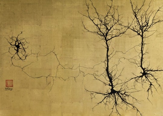
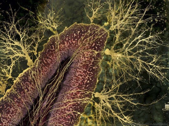
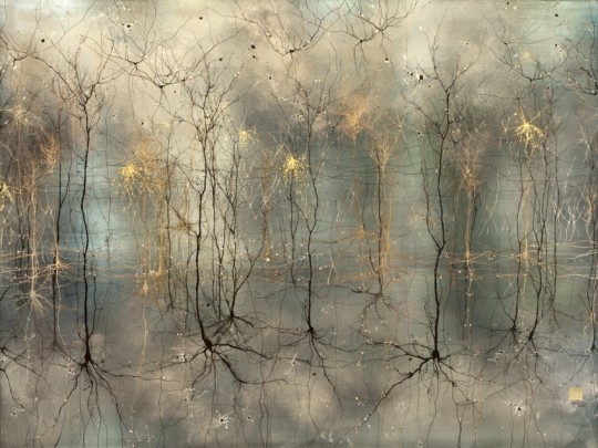
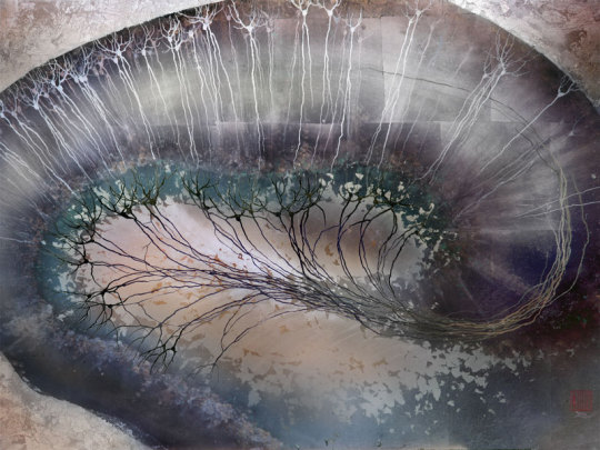
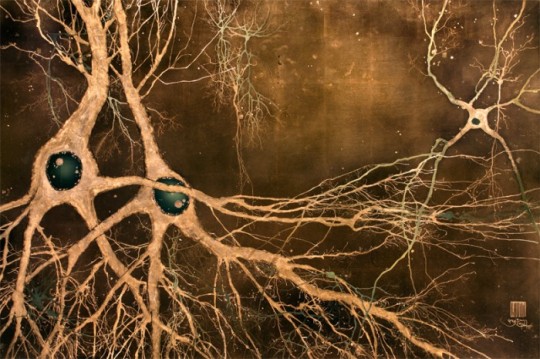
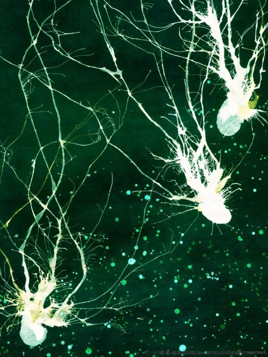
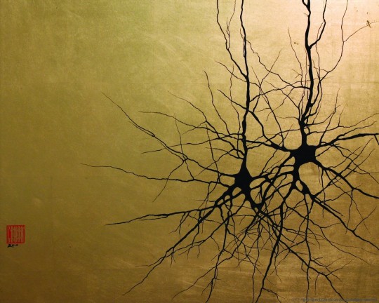
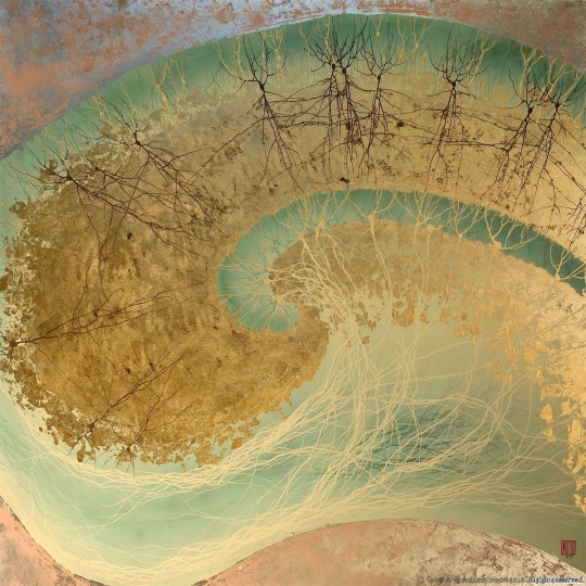
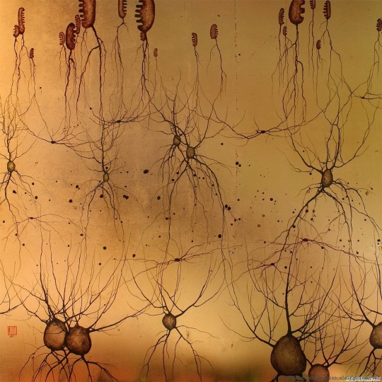
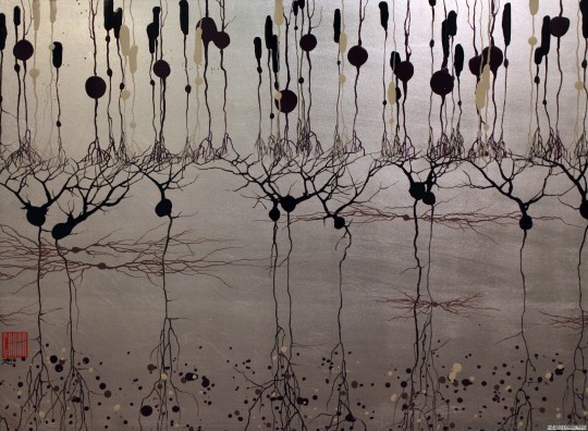
These amazing landscapes of the brain in gold, ink, dye, and metal, are by neuroscientist-artist Greg Dunn , who is inspired by the sumi-e style of ink wash painting. He also uses the technique of microetching, a form of lithography that manipulates light on a microscopic scale, which creates a very beautiful and stunning effect.
1K notes
·
View notes
Photo
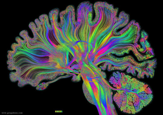
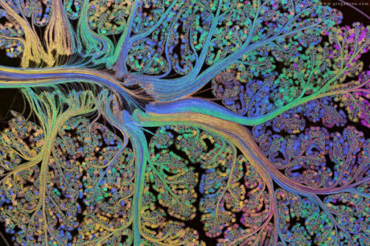
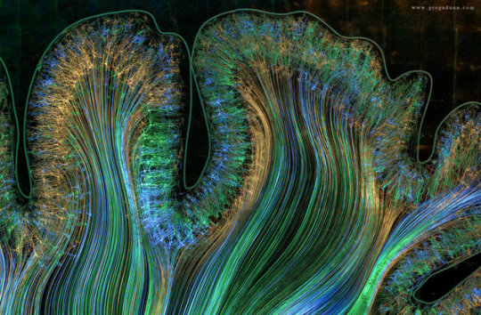
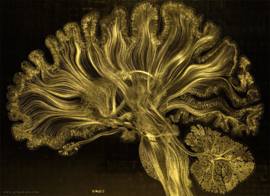
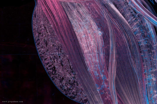

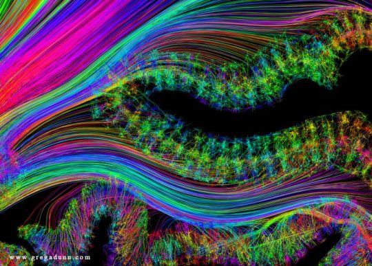
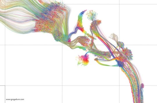
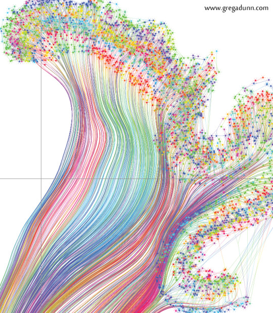
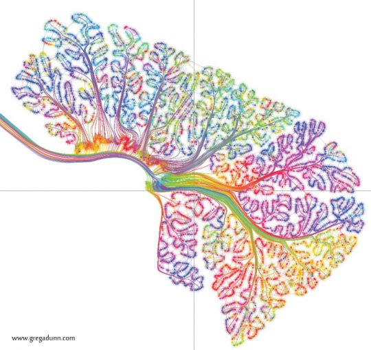
Giant Artwork Reflects The Gorgeous Complexity of The Human Brain
The new work at The Franklin Institute may be the most complex and detailed artistic depiction of the brain ever.
Your brain has approximately 86 billion neurons joined together through some 100 trillion connections, giving rise to a complex biological machine capable of pulling off amazing feats. Yet it’s difficult to truly grasp the sophistication of this interconnected web of cells.
Now, a new work of art based on actual scientific data provides a glimpse into this complexity.
The 8-by-12-foot gold panel, depicting a sagittal slice of the human brain, blends hand drawing and multiple human brain datasets from several universities. The work was created by Greg Dunn, a neuroscientist-turned-artist, and Brian Edwards, a physicist at the University of Pennsylvania, and goes on display at The Franklin Institute in Philadelphia.
“The human brain is insanely complicated,” Dunn said. “Rather than being told that your brain has 80 billion neurons, you can see with your own eyes what the activity of 500,000 of them looks like, and that has a much greater capacity to make an emotional impact than does a factoid in a book someplace.”
To reflect the neural activity within the brain, Dunn and Edwards have developed a technique called micro-etching: They paint the neurons by making microscopic ridges on a reflective sheet in such a way that they catch and reflect light from certain angles. When the light source moves in relation to the gold panel, the image appears to be animated, as if waves of activity are sweeping through it.
First, the visual cortex at the back of the brain lights up, then light propagates to the rest of the brain, gleaming and dimming in various regions — just as neurons would signal inside a real brain when you look at a piece of art.
That’s the idea behind the name of Dunn and Edwards’ piece: “Self Reflected.” It’s basically an animated painting of your brain perceiving itself in an animated painting.
To make the artwork resemble a real brain as closely as possible, the artists used actual MRI scans and human brain maps, but the datasets were not detailed enough. “There were a lot of holes to fill in,” Dunn said. Several students working with the duo explored scientific literature to figure out what types of neurons are in a given brain region, what they look like and what they are connected to. Then the artists drew each neuron.
Dunn and Edwards then used data from DTI scans — a special type of imaging that maps bundles of white matter connecting different regions of the brain. This completed the picture, and the results were scanned into a computer. Using photolithography, the artists etched the image onto a panel covered with gold leaf.
“A lot of times in science and engineering, we take a complex object and distill it down to its bare essential components, and study that component really well” Edwards said. But when it comes to the brain, understanding one neuron is very different from understanding how billions of neurons work together and give rise to consciousness.
“Of course, we can’t explain consciousness through an art piece, but we can give a sense of the fact that it is more complicated than just a few neurons,” he added.
The artists hope their work will inspire people, even professional neuroscientists, “to take a moment and remember that our brains are absolutely insanely beautiful and they are buzzing with activity every instant of our lives,” Dunn said. “Everybody takes it for granted, but we have, at the very core of our being, the most complex machine in the entire universe.”
Image 1: A computer image of “Self Reflected,” an etching of a human brain created by artists Greg Dunn and Brian Edwards.
Image 2: A close-up of the cerebellum in the finished work.
Image 3: A close-up of the motor cortex in the finished work.
Image 4: This is what “Self Reflected” looks like when it’s illuminated with all white light.
Image 5: Pons and brainstem close up.
Image 6: Putkinje neurons - color encodes reflective position in microetching.
Image 7: Primary visual cortex in the calcarine fissure.
Image 8: Basal ganglia and connected circuitry.
Image 9: Parietal cortex.
Image 10: Cerebellum.
Credit for all Images: Greg Dunn - “Self Reflected”
Source: The Huffington Post (by Bahar Gholipour)
6K notes
·
View notes
Photo


Watch: 12-year-old Arturo also explains to anti-vaxxers why it’s not “my child, my choice.”
Follow @the-future-now
82K notes
·
View notes
Photo

First structural views of the NMDA receptor in action will aid drug development
Structural biologists at Cold Spring Harbor Laboratory (CSHL) and Janelia Research Campus/HHMI, have obtained snapshots of the activation of an important type of brain-cell receptor. Dysfunction of the receptor has been implicated in a range of neurological illnesses, including Alzheimer’s disease, Parkinson’s disease, depression, seizure, schizophrenia, autism, and injuries related to stroke.
Led by CSHL Associate Professor Hiro Furukawa, the research team has obtained images of the NMDA (N-methyl, D-aspartate) receptor in active, non-active, and inhibited states. Understanding how NMDA receptors activate is critical in designing novel therapeutic compounds. NMDA receptors are embedded in the membrane of many nerve cells in the brain and are involved in signaling between cells that is essential to basic brain functions, including learning and memory formation.
Structurally, the NMDA receptor is made up of various protein segments, called domains, which together resemble a hot air balloon. The upper, balloon-like portion is comprised of the amino terminal domain (ATD); protruding from the outer surface of the cell is the ligand binding domain (LBD); and the lower, basket-like portion of the receptor, called the transmembrane domain (TMD), drops down inside the cell.
Activation of the NMDA receptor requires binding of brain chemicals called neurotransmitters at specific sites on the LBD. This binding together with structural rearrangement of the ATD triggers the opening of the channel formed by the TMD. This molecular event causes charged atoms called ions flow into the cell. When this occurs in many channels at once, an electrical current is generated that rapidly propagates through the neuron and triggers the release of neurotransmitters. These chemical signals, in turn, bind to receptors on neighboring cells.
Despite accumulating knowledge regarding the structure of the NMDA receptor and its various components, precise, details about the structural movements leading to the process of opening of the ion channel have not been described previously and the mechanism of receptor activation has remained unclear.
In addition to describing the NMDA receptor’s balloon-like structure, Furukawa and the team of CSHL structural biologists have previously revealed many important features of NMDA receptors, including the distinctive ways in which a number of drug compounds attach to the receptor at its various binding sites. “The NMDA receptor architecture itself is quite complicated,” says Furukawa, “but most recently we’ve been really fascinated by how each of its domains moves in a sophisticated but organized manner.”
To learn more about the dynamics of NMDA receptor activation, the researchers combined two molecular imaging techniques, x-ray crystallography and single-particle electron cryomicroscopy, and observed structures of the NMDA receptor in three specific configurations, the activated, non-active, and inhibited states.
Each of these configurations was achieved by the binding of different molecules to the receptor. For example, the activated state required the binding of the neurotransmitters glycine and glutamate, while the binding of the compound ifenprodil resulted in the inhibited state that forces the channel to be closed. The researchers used these molecular structures, published in Nature, to infer how the receptor’s various subdomains rearrange themselves when transitioning from a non-active to the active state.
“With the technology available today, we don’t see continuous movement but instead we see snapshots of NMDA receptors in different functional states,” Furukawa notes.
Superimposing the crystal structure of the NMDA receptor in each of the three functional states revealed which components move—typically by rotating slightly relative to one another—when the ion channel opens. The researchers observed that activation required opening of the bi-lobed architecture of the one of the two ATD subdomains and a reorientation of the ATD as a whole. These changes lead to additional rotations at various points throughout both the ATD and LBD, causing the ion channel pore to open, similar to the opening of a camera shutter.
Furukawa says that studying how components of the NMDA receptor move during activation and inactivation can help scientists create computer simulations to predict how the structures of various drug molecules might impact these movements. “Unless we have a library of molecular structures, people in the field won’t be able to run those simulations,” says Furukawa. “We hope that this new finding will help pharmacologists come up with better therapeutic compounds that have minimal side effects.”
185 notes
·
View notes
Photo

Horsehead Nebula Blue to Infrared (desktop/laptop) Click the image to download the correct size for your desktop or laptop in high resolution
99 notes
·
View notes
Photo
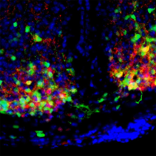
Mapping the circuit of our internal clock
What’s that old saying about Mussolini? Say what you will but he made the trains run on time. Well, the suprachiasmatic nucleus — SCN for short — makes everything in the body run on time. The SCN is the control center for our internal genetic clock, the circadian rhythms which regulate everything from sleep to hunger, insulin sensitivity, hormone levels, body temperature, cell cycles and more.
The SCN has been studied extensively but the underlying structure of its neural network has remained a mystery.
Now, researchers from the Harvard John A. Paulson School of Engineering and Applied Sciences (SEAS), the University of California Santa Barbara, and Washington University in St. Louis have shown for the first time how neurons in the SCN are connected to each other, shedding light on this vital area of the brain. Understanding this structure — and how it responds to disruption — is important for tackling illnesses like diabetes and posttraumatic stress disorder. The scientists have also found that disruption to these rhythms such as shifts in work schedules or blue light exposure at night can negatively impact overall health.
The research was recently published in the Proceedings of the National Academies of Science (PNAS).
“The SCN has been so challenging to understand because the cells within it are incredibly noisy,” said John Abel, first author of the paper and graduate student at SEAS. “There are more than 20,000 neurons in the SCN, each of which not only generates their own autonomous circadian oscillations but also communicates with other neurons to maintain stable phase lengths and relationships. We were able to cut through that noise and figure out which cells share information with each other.”
The SCN looks like a miniature brain, with two hemispheres, inside the hypothalamus. It receives light cues from the retina to help it keep track of time and reset when necessary. When functioning properly, the neurons inside both hemispheres oscillate in a synchronized pattern.
In order to understand the structure of the network, Abel and the team had to disrupt that pattern. The researchers used a potent neurotoxin commonly found in pufferfish to desynchronize the neurons in each hemisphere, turning the steady, rhythmic pulse of oscillations into a cacophony of disconnected beats. The team then removed the toxin and observed the network as it reestablished communication, using information theory to figure out which cells had to communicate to resynchronize the whole network.
“It’s like trying to figure out if a group of people are friends without being able to look at their phone calls or their text messages,” Abel said. “In a large group of other people, you might not be able to tell who is in contact with each other, but if a certain group shows up together at a party, you can probably assume they’re friends because they show similar behavior.”
By observing the SCN at single-cell resolution, Abel and the team identified a core group of very friendly neurons in the center of each hemisphere that share a lot of information during resynchronization. They also observed dense connections between the hubs of each hemisphere. The neurons outside these central hubs, in the area called the shell, behaved more like acquaintances than friends, sharing little information amongst themselves.
“We were surprised to find that the shell lacked a functionally connected cluster of neurons,” said Abel. “We’ve known that exposure to an artificially long day can split the SCN into core and shell phase clusters which oscillate out of sync with each other. We’ve assumed that the neurons in the shell communicated to synchronize that rhythm but our research suggests that phase clustering in the shell is actually mediated by the core neurons.”
Previous research also assumed that the core SCN was dominant only due to its role in receiving light cues from the eyes. By using the neurotoxin to disrupt circadian rhythms, Abel and the team demonstrated that the core is the key to resynchronization even without light cues.
“For the last 15 years our group has been studying the complex control mechanisms that are responsible for the generation of robust circadian rhythms in the brain,” said Frank Doyle, the John A. Paulson Dean and John A. & Elizabeth S. Armstrong Professor of Engineering & Applied Sciences, who co-authored the paper. “This work brings us one step closer to reverse engineering those paradigms by elucidating the topology of communication amongst neurons, thus demonstrating the importance of a systems perspective to link genes to cells to the SCN tissue.”
108 notes
·
View notes
Photo

Orion Nebula (phone) Click the image to download the correct size for your phone in high resolution
145 notes
·
View notes
Photo


How to create a (realistic) fictional language
M'athchomaroon. That’s a hello to you, in Dothraki.
Initially, it may be easy to dismiss those words from the fictional language in “Game of Thrones” as a bunch of made up gibberish, but upon closer inspection, you might realize that the speech and word patterns resemble a real language.
And that’s because it is, in fact, a language with its own fully functional grammar and over 4,000 words.
Before you can even begin writing a single word in Dothraki, you have to do a ton of foundational work to make the constructed language (conlang) seem authentic and natural.
“I used the books almost as anthropological text. Paying attention not just to the dialogue in any given chapter, but also the description of what the land was like, and what people were doing, what they were eating, and wearing,” says David Peterson, the creator of the Dothraki dialect and a UC San Diego alum.
All this detailed analysis of the characters’ realities, culture and attitudes informed the words that would exist in that language.
Here’s an example: Since the Dothrakis are nomadic warriors who believe in taking what they want through brute force, there is no word for “thank you.” But there are seven words just for swinging a sword (like “hlizifikh,” which is a wild, but powerful strike.)
And horse riding is so entrenched in their culture, that their very name Dothraki is derived from their verb “to ride”: dothralat.
Just as modern English was developed from its Old English form, Peterson also created an antiquated version of Dothraki and a modern version. Like real languages that have existed, each word has an etymology that reflects how the language evolved over time.
All this may seem like an insane amount of work and thought for a few lines of dialogue, but for a language enthusiast like Peterson —who speaks eight languages— creating a conlang is a self-indulgent hobby. It’s fun.
“Creating a language is an art form, like any other. I enjoy doing it. I don’t really think about the endpoint… after all, a language is never really finished,” says Peterson, who would continue to conlangle even if he wasn’t getting paid.
For budding conlangers, Peterson recently developed some resources, including a book and YouTube series that teach more about the process of inventing languages and the history of conlang in further detail.
@teded also has this great video all about fictional languages:
youtube
GIF: TedEd
2K notes
·
View notes
Video
youtube
Jumping electric eels pack more zap
To take the zap out of a school of electric eels, fishermen in 17th century South America sent teams of horses into the water as bait, scooping up the eels after they had exhausted themselves in the attack. According to famed naturalist Alexander von Humboldt, the eels would leap out of the water to shock the frightened—but mostly unharmed—horses. Until now, no one else had recorded evidence of such behavior, and many scientists were skeptical of Von Humboldt’s account. A new study shows that not only do the animals leap from the water to attack their prey, but they also increase their voltage as they leap (see video). The jumping behavior was first observed by accident: As scientists were studying the eels in an unrelated experiment, they noticed that the animals would sometimes jump to attack the rim of the nets used to capture them. In a follow-up study, reported today in the Proceedings of the National Academy of Sciences, the researchers found that the attacks became markedly more powerful the higher the eel rose out of the water, in one case increasing from 10 to 300 volts. As the animal leaves the water, electrical resistance through its body increases, forcing the charge into a less resistant target—its prey. The farther the eels can get out of the water, the more current they can deliver—a shocking bit of evidence to support Von Humboldt’s electric account.
81 notes
·
View notes

