#surgical peritonitis
Explore tagged Tumblr posts
Visit Tumblr Blog
Explore Tumblr blogs with no restrictions, modern design and the best experience.
Last Seen Tumblr Blogs


Fun Fact
Tumblr has been banned in Indonesia for providing people with access to pornographic content.
Text
Let's Talk ESRD and Dialysis
Have you thanked your kidneys today? Do you feel grateful when you pee? How about when you eat a little too much potassium or drink a little too much water, do you really enjoy feeling confident that your kidneys will just dispose of the excess?
If so, you probably know the alternative.

About 10% of the world's population has a condition called Chronic Kidney Disease, or CKD. About 2 million of those people are in End Stage Renal Disease (ESRD) and require dialysis or a kidney transplant to live.
Your kidneys are amazing things. They are two organs that sit outside of the sac that hold the rest of the abdominal organs, called the peritoneum. They take in blood from the body, determine the levels of electrolytes, water, and waste products in that blood, and remove the waste products and excess electrolytes and water.
They also have secondary tasks. They monitor the amount of red blood cells in your blood and send out hormones that entice the bone marrow to make more when we're low. They also monitor blood pressure and release hormones that raise that blood pressure when it gets low.
Lots of things can hurt the kidneys. For example, poorly controlled high blood pressure and poorly controlled diabetes are among the top reasons why kidneys fail. Additionally, being dehydrated while engaging in strenuous exercise or taking medications like ibuprofen or naproxen (any NSAIDs) can cause kidney damage.
We measure how well the kidneys are working via the Glomerular Filtration Rate, or GFR. This is a measure of (essentially) how much blood in milliliters the kidneys filter per minute. 90 or higher is normal, while a GFR of 15 or lower is considered ESRD.
So let's say someone has a GFR of less than 15 and the decision is made to start them on dialysis and put them on the kidney transplant list. What options do they have?
Well, they need to figure out if they want to do hemodialysis or peritoneal dialysis.
In hemodialysis, the patient is hooked up to a machine that runs their blood across a special membrane. On the other side of the membrane, a solution called dialysate draws excess water, electrolytes, and waste products from the blood. Hemodialysis is usually done at a dialysis center for 3-5 hours, 3 times per week.

Hemodialysis is better for patients who have either failed home peritoneal dialysis or can't or aren't comfortable with doing the technical part of the job by themself at home. There is also a social component, where dialysis is a chance to meet and interact with other people who are going through the same things they are.
People who undergo hemodialysis have to have some kind of "access", or a way for the blood to come out of their body, go through a machine, and go back into their body. For some people, this is a dialysis catheter that is inserted into the person's chest and looks like this:

It can also be a fistula. A fistula is the surgical connection between a vein and an artery in the arm or leg. Over time, this connection becomes large and rubbery, and each time dialysis is done, two needles (one to remove blood, and one to return it) are placed in the fistula. A fistula often looks like this:

In peritoneal dialysis, the patient instills the dialysate directly into the sac that holds their abdominal organs. The sac itself acts as the membrane, and dialysate draws the electrolytes, water, and waste directly through the sac wall. They then wait a certain number of hours, and drain the dialysate. This can be done manually by the patient during the day, or at night while the patient sleeps with a machine called an automatic cycler. Usually peritoneal dialysis is done every day, with 2-4 cycles of 4 hours per cycle.
People using peritoneal dialysis also need a form of access, but instead of it being to their blood stream, it is to their peritoneum. Here's what that looks like:

The catheter is placed surgically into the peritoneum, and stays there all the time, even in between dialysis sessions.
Someone using peritoneal dialysis has to be very careful when they are accessing their dialysis catheter. This is because the biggest problem with peritoneal dialysis is the risk of a life threatening infection called peritonitis. Someone who gets peritonitis too many times may need to switch to hemodialysis.
Here is what a manual exchange looks ilke:
youtube
Someone may choose to do peritoneal dialysis over hemodialysis because it affords more freedom to keep a job or do daily tasks like keeping house. People who do PD also don't have to find rides to the dialysis center. However, they do have to take on more of the responsibility for making sure they do treatments correctly and be able to keep accurate records of the treatments they give themselves. Peritoneal dialysis also tends to be less taxing on the body, and have fewer side effects than hemodialysis when done correctly.
36 notes
·
View notes
Text
the worst thing about love is… | jason todd

the worst thing about love
you’re just trying to get through your surgical residency, but this masked vigilante keeps showing up half-dead on your fire escape and reminding you of your dead best friend. oh well, at least he's cute.
one | two | three | series masterlist
tw: stitches, mentions of blood and injuries, swearing, completely ooc Jason but he’s like my own lil character now and I’m protective, very inaccurate medical terminology and procedure lol
only jerks steal other people's writing (just don't repost, mate)
There’s a dead man on your fire escape.
Well. He’s not actually dead, but his pulse is weak when you drag him into your living room, out of the relentless Gotham rain. Pulling your hand away from under his mask, you crouch down, peeling off the worn leather jacket around his shoulders and unbuckling his pauldrons. You feel around his back, brows furrowed. You can’t feel anything through the padding in his rain soaked shirt.
Hands wandering down to where his front is flat on the floor, you press down on his side, eyes widening when your fingers come back slick with blood. You go into autopilot, flipping him onto his back and yanking up his compression shirt. You might’ve gasped at the knife wound if you weren’t working on instinct. It’s bad.
Shoving away the doubt clawing at the base of your skull, you steady your trembling hands. You’ve been trained for this.
Don’t feel, just do.
The cut is long and serrated, and deep as all hell. It slices through the middle of a jagged, Y-shaped scar that chains over his shoulders like a noose. Jesus.
It’s like he was stabbed and then dragged across the floor, cutting diagonally across his torso. How is he even still alive? Your hands move faster than you can think, completing an internal checklist as you go.
Breathing? Fast and shallow through his modulator, no obstructions. Bleeding? Applied tourniquet to epigastric region - transfusion isn’t even an option… Your brain works overtime, sifting through diagnostics lectures - penetrating abdominal trauma, debrided of devitalised tissue, no visible debris… You trace the edges of the wound looking for inflammation or fluid buildup; signs of peritonitis, but the weapon seems to have missed any internal organs. Lucky. Even luckier that he landed on a surgical resident’s fire escape.
Reaching over to the lamp by your couch, you shift it so that it shines directly over his abdomen. A last check of his wound confirms that there are no external indications that you should conduct a laparotomy. You just have to sew him up and hope to god the knife didn’t puncture anything internal.
You keep a hand planted firmly over his tourniquet, applying constant pressure, reaching for your backpack. Dragging it over, you use your teeth to open your suture kit and your free hand to sterilise his cut with Betadine and alcohol, wiping gentle circles outward from the wound. You dip your needle like Achilles in the Styx, hand and all, into the sterilising liquid, tugging a glove on with your teeth.
You grip the needle driver in your dominant hand, pickups clutched in the other and take a steadying breath. There’s a stillness to the room, quiet save for your heartbeat pounding in your ears. The wound is large - high tension - so… mattress sutures… horizontal so the tension is spread over the edge of the wound.
You take your first bite, adrenaline driving your needle into a clean stitch. You reverse it, passing through his cut again, before tying it off with the practised motions of a thousand surgical knots tied on yarn and thread and fraying jeans. You settle back on your knees after the first suture, readying yourself for the stitching to come, and start the next one.
~
Hours later, you haul him onto your couch, sitting him up on the arm rest to take pressure off of his dressed stitches. Frowning deeply at how uncomfortable he looks - even unconscious, you tuck a throw pillow under his scuffed metal mask.
Leaning close to check his breathing, you hear crackling slow and deep through the helmet’s voice modulator. Bone-deep relief floods your system, a little sigh leaving your mouth involuntarily. Sitting heavily against your coffee table, you press the heels of your hands into your weary eyes.
He’s stable. For now at least.
Head bumping against the edge of your couch, you breathe in deeply, fighting the anxiety twisting in your ribcage. The couch smells like rubbing alcohol, stinging your nose so badly your eyes water. It’s followed by something familiar - underneath the heady scent of petrol and metal - like… if you mixed Gotham up into a single smell; rain and smoke and wet pavement. He… he smells like-
“Jay!”
The faulty fluorescent lights - courtesy of your parent's small family diner - seem to flicker in tandem with your strident yell.
Your best friend looks up at you through a mop of dark hair, collarbones poking out of his thin t-shirt, second-hand leather jacket chucked haphazardly on the other side of the booth. He’s stolen your copy of Jane Eyre, flattened with one hand next to a plate of old fries you’d scrounged for him.
You tug your book from his grasp, tucking your pen into the pocket on your apron. He looks up at you with a mouth full of fries, infuriating confusion written across his face.
“What? You promised I could read it.” You sigh in exasperation.
“When I’m finished! And-” A dramatic gasp rips from your mouth when you examine the book. “Are these- grease stains?” You take the book in both hands, swatting Jason with it.
“What so it’s okay to hit me with a book but not get grease- fuck, jesus, okay, okay!” You raise the book over your shoulder with both hands.
“Do you yield?” His mock-angry expression almost makes you laugh, a hand held up near his face to shield from your attack. There’s a soft twist to his frown, like he’s trying to stop his mouth from pulling into a grin. He raises his hands in surrender, and you relax your hold on the book.
Rookie mistake.
Jason darts forward, faster than you can blink, grasping your waist with both hands and dragging you towards him. He yanks the book from your hands and lets you go, grinning childishly at you with the book in his hands. The cat with the canary.
You throw your hands up in exasperation before planting them on your hips like a disappointed mother. The admonishment on the tip of your tongue turns into a weary sigh when you hear your parents calling for you from the diner kitchen. “Fine. But you actually have to try to not spoil it this time.”
Jason crosses his fingers over his chest, “Scout’s honour, birdie.”
You try not to flush at the nickname, just like you do every time he says it. Still, you fold like a stack of cards.
(He spoils it the next day.)
~
When you wake two hours later for rounds (at the ass-crack of dawn), he’s already gone. You pad quietly around your kitchen making coffee from day-old grounds, cautious not to disturb the sanctity of the early morning (or the ghost of his presence).
The only evidence of him is alight in the dim light that spills over your kitchen counter and into your living room - the deep indents in your couch and the bloodstains on your carpet… The rain on your wood floors, from the fire escape window you’re sure you didn’t leave open.
hi, hello, uhh this is the first fic I've ever posted so bear with me. if anyone actually sees this, i do apologise for the inaccuracies and lengthy prose. also, this will be a series so stick around if you like slow updates, slowburn and second chances. thanks for reading my rambles.
with love, bugsy
#series: the worst thing about love#love-bugsy#jason todd x reader#jason todd fic#jason todd fanfiction#jason todd reader insert#jason todd imagine#x reader
250 notes
·
View notes
Text
‘Verse: Annihilation
Project Tempest Archives Footage : ██/██/███ – autopsy of P█████ A██████
A body lies naked on a steel table. Female, middle-aged, and a little overweight. The skin is blotched with inflammation and bruising, and dotted with innumerable tiny scabs. The abdomen is distended. Several surgical incisions mark the torso – recent and unhealed, but neatly closed with rows of sutures.
Masked and gloved surgeons step into the camera’s field of view, and take up tools. A similarly masked cameraperson joins them, holding a handheld camera.
They begin by opening an abdominal surgical site. Sutures are cut away from the surface of the skin, then from the layer below, and finally from the fascia. As the flesh parts, thick, clotted blood wells up from beneath, oozing like custard through the opening. The doctors wipe away the gobbets, and continue.
Scalpels lengthen the incision while retractors pull the sides of the wound back to expose the organs beneath. Close-up views alternate with the top down footage as the cameraperson moves between the surgeons with practiced ease.
The abdominal cavity is full of clotted blood. The doctors wipe it out by the gelatinous handful, discarding it as they proceed. The close up shots offer little to the untrained eye but meat, even as the surgeons use forceps to indicate certain bruised tissues and sutured vessels.
Once done with the first surgical site, the team moves onto the next, and the next. After each has been inspected in turn, the surgeons open the abdominal cavity from sternum to pelvis and begin removing organs. More congealed blood is scooped away.
The close up camera focuses on each organ in turn. An abraded surface here, a dark bruise there. The stomach is emptied of its contents – more dark blood.
They saw open the ribcage to access heart and lungs. Clots spill from the thoracic cavity. The lungs themselves are blotched with the deep red of blood. The heart is intact, coated with streaks of yellow fat.
Finally, they open the cranium and extract the brain, dissecting it beneath the unflinching eye of the camera to point out the sites of multiple bleeds.
The footage ends abruptly, without outro or closing remark.
---
Attached is the full autopsy. The primary cause of death is determined to be pulmonary haemorrhage. A long list follows of the other sites of haemorrhage.
Systemic diffuse tissue damage is noted, as are high levels of systemic inflammation. Early signs of infection were found in the blood-filled peritoneal cavity. Acute kidney failure was noted shortly before death.
Multiple surgeries were performed before the subject’s death, targeting the sites of the most severe bleeding. Full notes are attached after the autopsy.
15 notes
·
View notes
Note
what would be the affects of a cut wound to the side of the abdomen, about five inches long, one inch deep, that was left for an hour with the only care to it being pressure applied with a rag?
Depends on if the cut entered the abdominal cavity or just cut through fat and muscle.
If the cut was just through fat and muscle, not much. It would bleed but not life-threateningly, and would need cleaned well and either packed (remote/wilderness setting) or closed surgically (hospital setting). Antibiotics and a tetanus shot would probably be given to prevent infection.
If the cut was into the abdominal cavity (the sac that holds the abdominal organs), the bleeding again wouldn't be life threatening, but now you're talking an actual need for a very specific kind of cleaning and surgery, and probably IV antibiotics to prevent a serious infection called peritonitis.
77 notes
·
View notes
Text
“Sophia Roe,” 19 (USA 1969)
The unusual aspect of “Sophia’s” death is that although the abortion that killed her was unambiguously illegal at the time, it was declared “retroactively legal” due to Roe v. Wade and her killer was released from prison.
“Sophia” was a college student at Ohio State University. The illegal abortionist Benjamin King was known to circulate his contacts on college campuses to lure vulnerable pregnant students to him. When Sophia and her boyfriend (who will be referred to here as “Oliver”) found out she was pregnant, they made the mistake of trying out this shady contact for what was at the time an illegal abortion.
Sophia and Oliver, both 19, made a trip from Youngstown, Ohio to Duquesne, Pennsylvania just like King told them to on December 27, 1969. They paid $300 (over two thousand in today’s money) as a down payment. King proceeded with the surgical abortion that would end two lives.
The young couple made their way back to school after the abortion, but it rapidly became obvious that something was very wrong with Sophia. She was admitted to South Side Hospital on December 29, but died the next day. It was discovered that King caused a perforated cervix, peritonitis, massive hemorrhage and shock.
Oliver worked undercover with police to catch his girlfriend’s killer. He contacted King and told him that he had the rest of the money. When King came to collect it, he was arrested for Sophia and her child’s murder. Back then, the state’s law stated that death of a pregnant woman from abortion qualified as murder. It looked like there would be some semblance of justice for Sophia and her baby.
But Roe v. Wade deprived Sophia and her loved ones of justice. When abortion was legalized, King managed to make the argument that the abortion that killed Sophia and the baby was “retroactively legal,” which a court approved. Thanks to Roe v. Wade, Sophia’s fatal abortion was now classified as legal and her murderer walked free, now able to endanger other pregnant women without the fear of prosecution for their deaths.
Sophia’s death is similar to the case of Nancy Ward in 1968 and Sara Carr in 1965. Nancy’s killer and Sara’s killer were also released after a ruling from Roe v. Wade made the fatal abortions retroactively legal.
#tw abortion#pro life#pre roe legal#retroactively legal#unsafe yet legal#tw ab*rtion#tw murder#abortion#abortion debate#death from legal abortion#unidentified victim
14 notes
·
View notes
Note
Our favorite dramatic medical emergency that can be fixed quickly, appendicitis? How can it go wrong and that should people expect in the hospital?
Ah, appendicitis... A classic and personal favorite of mine. Most of my poor whumpees have had to endure this one.
Acute appendicitis initially presents with generalized abdominal pain that migrates to the lower right abdomen, decreased appetite, nausea and vomiting, diarrhea, malaise, and decreased urinary output. A fever may be present. Person will likely be guarding the abdomen and putting their knees to their chest to relieve pressure. The hallmark sign of appendicitis is the McBurney sign: the person experiences a reduction in pain when someone presses on their lower right abdomen, but their pain increases when the pressure is removed (rebound tenderness).
The most common complication of appendicitis is rupture and resultant peritonitis (inflammation of the lining of the abdominal cavity [peritoneum]). Rupture can occur as early as 24 to 48 hours after the onset of symptoms, but may take longer (risk of rupture increases by 10% with each passing day). Despite how it sounds, rupture is usually not painful and may even bring temporary relief due to the release of pressure. However, fecal matter from the intestines is now leaking freely into the abdominal cavity and peritonitis can set in in 24 hours. Peritonitis presents with fever, chills, abdominal pain, diarrhea, and obstipation (inability to pass gas or stool). The hallmark sign is board-like abdominal rigidity.
Appendicitis without rupture is managed with an emergency laparoscopic appendectomy and antibiotics. Rupture with or without peritonitis is treated in the same way, though an open appendectomy may be performed if extensive contamination or abscesses are present. Peritonitis requires a longer antibiotic regimen than simple appendicitis.
Recovery after an appendectomy is pretty standard postoperative recovery. The patient will likely be in the medical-surgical unit, but peritonitis might warrant an ICU admission. This post describes postop recovery pretty comprehensively.
The patient will initially have a urinary catheter, but this will be removed as soon as the patient can safely walk to the bathroom.
The patient will be NPO (nothing by mouth) until bowel sounds are present in all four abdominal quadrants and the patient has passed gas or stool, after which they will be put on clear liquids, and then solid food if those are tolerated well.
The patient will be encourage to stand, sit in a chair, and walk with support as soon as possible. While in bed, the patient will have pneumatic compression devices on their legs that will intermittently squeeze their legs to keep the blood flowing.
The patient will be encouraged to use an incentive spirometer at least once per hour and cough and deep breath frequently.
The medications that the patient will receive include around-the-clock opioids, blood thinners, stool softeners, and antibiotics.
The patient with uncomplicated appendicitis while likely be discharged within 24 hours of surgery, but a patient with peritonitis may stay in the hospital for a few days and up to a week. Staples will be removed and replaced with Steri-strips 1 to 2 weeks after surgery, and the patient should be fully recovered in 2 (for an uncomplicated appendectomy) to 6 weeks (for an open appendectomy or peritonitis).
Happy whumping!
4 notes
·
View notes
Text
Kasotakis, G., Roediger, L., & Mittal, S. (2011). Rectal foreign bodies: A case report and review of the literature. International Journal of Surgery Case Reports, 3(3), 111–115. https://doi.org/10.1016/j.ijscr.2011.11.007
available here
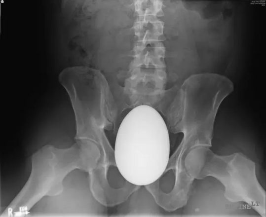
A 41-year-old HIV+ Caucasian male presented to the emergency department (ER) complaining of severe pelvic pain from a large oval-shaped marble he had inserted in his rectum approximately 2 h prior to presentation. The patient reported that multiple attempts to remove it at home failed, even with use of marijuana (in an effort to relax the anal sphincter) prior to his arrival at the ER.
On examination, his abdomen was soft, non-distended and non-tender to palpation, without sings of peritonitis. Bowel sounds were decreased. An X-ray of the lower abdomen revealed a large, ovoid-shaped object in the rectum (Fig. 1). The foreign body was palpable in the rectum, but due to its shape, large size and its smooth surface it was impossible to retrieve with simple maneuvering, including simultaneous application of suprapubic pressure. Proctoscopy was not attempted, as the anal canal was well dilated and the foreign object and distal rectal mucosa were easily seen and examined with a rectal speculum. Mild mucosal hyperemia was noted, but there was no evidence of tears or ischemic compromise to the rectal mucosa. As the patient was very uncomfortable with our maneuvers, despite maximal intravenous analgesia, we elected to proceed with an examination under anesthesia and possibly surgical exploration.
After fluid resuscitation and preoperative intravenous antibiotics, the patient was brought to the operating room, where he was anesthetized and intubated, and placed in the lithotomy position. An attempt to remove the foreign body manually with lubrication and more aggressive manipulation was fruitless, as the foreign body's greatest diameter appeared to be wider than the patient's pelvic outlet. We attempted use of delivery forceps but were unsuccessful. A decision was made to proceed with laparotomy. We felt at attempt at laparoscopy would have been inadequate for extraction, given the size of the foreign item. An 8 cm midline incision was made infraumbilically and was deepened through the midline subcutaneous tissue and fascia with electrocautery, until the peritoneal cavity was entered. The distal sigmoid and rectum were identified and the foreign body was palpated below the pelvic brim, tightly wedged in the pelvis. It seemed that the marble was pushed into the rectum with force that transiently relaxed the pelvic ligaments and allowed its slightly wider diameter to pass through and wedge within the lesser pelvis. Unfortunately, due to the android shape of our patient's pelvis, we were unable to perform the same maneuver with downward force from the abdomen. As the proximal rectal wall was sliding over the apex of the foreign body, not allowing significant force to be applied uniformly onto it, and in order to prevent mucosal injury by compressing it against the foreign body with excessive pressure, an enterotomy was made through which the foreign object was again pushed downward toward the anus, again without results. An attempt at pushing the egg upward, from the rectum into the peritoneal cavity was similarly unsuccessful.
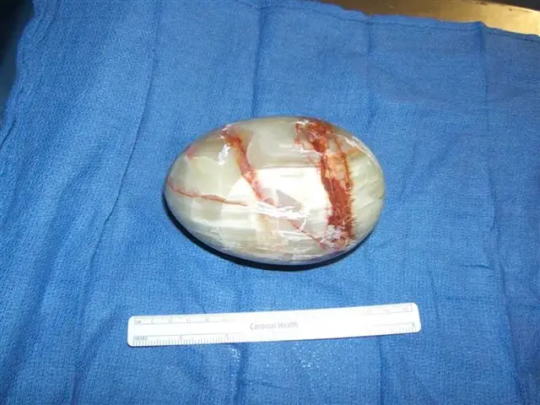
At this point we felt that it was the patient's pelvic anatomy that prevented us from retrieving the tightly wedged object and we consulted orthopedic surgery. A separate Pfannenstiel incision was made just over the superior edge of the pubis at the insertion of the rectus muscle. The incision was carried down through the subcutaneous tissue all the way down to the superior border of the symphysis. The dissection extended along the superior pubic rami in both directions laterally, the anterior and undersurface of the symphysis pubis anteriorly and posteriorly respectively, while care was taken to prevent bladder injury, transposing a protective wide malleable retractor between the urinary bladder and the pubic symphysis. The latter was divided longitudinally with an osteotome and stretched open to approximately 4 cm in width with a laminar spreader. Obstetric forceps were again used transanally to grasp the foreign body and pull it out, with the simultaneous application of downward manual pressure from the peritoneal cavity. The specimen, an egg-shaped, marble ornament measuring 12 cm × 8 cm × 8 cm, was sent to pathology for examination (Fig. 2).
Sigmoidoscopy was next undertaken and revealed minor mucosal bleeding over the areas that were compressed by the foreign body against the non-compliant bony pelvis. The enterotomy was closed with interrupted absorbable suture in two layers and checked with insufflation. After removal of the laminar spreader, a 1.5 cm gap remained at the symphysiotomy. No internal fixation implants were used due to contamination of our field from the enterotomy.
By this time, blood-tinged urine was noted in the Foley catheter, and bladder injury ruled out with intravesical irrigation followed with no evidence of extravasation, as the bladder was visualized through the opening in the symphysis pubis. The balloon of the urinary catheter was easily palpated and so was the prostate. Cystoscopy was deemed unnecessary due to absence of any obvious bladder injury on irrigation. No bleeding was noted from the venous plexus in the area and the Foley catheter was put to dependent drainage. Incisions were closed in layers.
The patient had an unremarkable recovery and was discharged on post-operative day 4 with some discomfort with ambulation.
2 notes
·
View notes
Text
Transcatheter arterial Embolization of the common hepatic artery for pseudoaneurysm after a laparoscopic-assisted pancreaticoduodenectomy: A case report by Yongxiang Li in Journal of Clinical Case Reports Medical Images and Health Sciences
Introduction
Pancreaticoduodenectomy (PD) is the main procedure for some surgeries related to the pancreas. Due to the advance of the surgical technology in recent two decades, mortality decreased considerably [1]. However, the morbidity rate for the major complication after PD remains high [2]. In the various complications, postpancreatectomy hemorrhage (PPH) is a fatal complication, which is linked with 11%−38% of the overall mortalities [3−6]. According to the International Study Group of Pancreatic Surgery [7], late PPH is caused by a ruptured pseudoaneurysm. Once the pseudoaneurysm ruptures, laparotomy and endovascular intervention are the main treatment to be done. Here we report the clinical features, diagnosis, and treatment of a case of massive hemorrhage in the common hepatic artery (CHA) for pseudoaneurysm after PD.
Case report
A 48-year-old male patient underwent a modified Child PD for the malignant tumor of the descending duodenum. The gastroscope and abdominal enhanced computed tomography (CT) in the preoperative examinations are displayed in Fig. 1. The related index and laboratory values of the patients showed no abnormal outcomes. Standard modified Child PD was performed after excluding the surgical contraindications. No adverse events occurred during the operation. Antibiotic prophylaxis was administered in the postoperative treatment. On postoperative day (POD) 2, the patient suffered from fever and abdominal pain. Persistent peritoneal lavage and drainage were conducted to prevent anastomotic leakage. On POD 8, the continuous drainage stopped because of disappearing abdominal pain. On POD 10, the patient had a sudden abdominal pain and showed 50 mL loss of blood from the drain of cholangiojejunostomy. Hemoglobin concentration decreased to 85 g/L, which had dropped by 45 g/L compared to the last inspection. At the same time, the amylase level measured in the intra-abdominal drainage fluid was 1480u/L. In terms of diagnosis, pancreatic fistula and intra-abdominal bleeding were considered. Conservative treatment, including fluid infusion, use of hemostatic agents, and blood transfusion, was used for this patient. Then, the patient’s condition was stabilized gradually. Abdominal CT was performed on the POD 19, which revealed the existence of bloody fluid collection around the perihepatic area (Fig. 2). On POD 21, the patient underwent catheter drainage under the guidance of ultrasonic from the perihepatic area. Abdominal distension of the patients improved. However, on POD 25, the patient abruptly developed melena and hematemesis, and vomited about 300 mL of bloody fluid. A total of 200 mL bright red bloody fluid drained from the abdominal tube. Then, the patient suffered from a shock with hypotension and tachycardia. Hence, Active abdominal bleeding was considered. Urgent Digital Subtraction Angiography (DSA) performed on the basis of a joint decision between the interventional radiologist and a surgeon. DSA revealed a pseudoaneurysm after the rupture of the CHA (Fig. 3a, Video 1). Then, embolization of the hepatic artery with microcoil was performed successfully (Fig. 3b, Video 2). The patient’s blood pressure returned to normal after embolization. And then the patient regained hemodynamic stability and was transferred to the Intensive Care Unit (ICU). The patient was successfully discharged from the hospital on POD 38. There were no obvious abnormalities in the patient’s reexamination after three months.
DSA procedure:
The patient lied supine on the DSA table; a puncture in the right femoral artery was performed after local anesthesia. The 5FRH catheter was placed into the right femoral artery, the catheter head was inserted into the celiac trunk artery for DSA, and the super-selected microcatheter (Terumo Progreat microcatheter, Japan) was inserted into the hepatic artery. After the hepatic artery, its branches were identified by contrast; the embolization microcoil was placed, followed by the injection of the histoacryl (B.Braun Closure Specialities, Germany) into the hepatic artery. Ultimately, the hepatic artery and its branches did not develop again and hence were not visualized under DSA.
Discussion
Commonly, complications develop after PD; there is no doubt that PPH is dangerous and fatal. Furthermore, a ruptured pseudoaneurysm is the most severe and fatal cause of PPH [8]. The formation of the pseudoaneurysm is associated with the damage to the vascular wall. Although adequate lymph node dissection and skeletonization of the vessels in surgery may significantly improve the patient’s prognosis, the dissection and skeletonization make the arterial wall weak and vulnerable, which is susceptible to erosion by trypsin and elastase from the digestive juice [9].
Then, we analyzed the pathogenesis of this case, which may be related to laparoscopic instrument operation. Especially, the dissociation of vessels and dissection of the lymph nodes caused excessive skeletonization, and then the Hem-o-lock ligation damaged the arterial wall, which may lead to the formation of the pseudoaneurysm in the stump of the ligated artery.
In this case, intraperitoneal hemorrhage occurred after surgery, and the measured drainage liquid amylase was 1480u/L; thus, it was considered that the digestive fluid leak caused by the pancreatic fistula, corroded the blood vessels and eventually led to bleeding. After conservative treatment, there is a possibility of hemodynamic instability that would require emergency DSA examination; the formation of a pseudoaneurysm of the CHA and arterial embolism are also considered. Microcoil was chosen given the hemodynamic instability of the patients; while the liver has a double blood supply, a simple embolism is not likely to cause liver ischemia necrosis. Microcoil and histoacryl embolization were chosen given.
A recent meta-analysis revealed that endovascular treatment of a ruptured pseudoaneurysm had low mortality and morbidity and high success rate than surgical intervention [10,11]. endovascular treatment is considered the first choice in the treatment of pseudoaneurysm recently. Endovascular treatment consists of Transcatheter Arterial Embolization (TAE) and stent-graft placement. Coil embolization as a TAE is an effective approach for the treatment of a pseudoaneurysm [12,13].
In this case, we summarized several experiences for the iatrogenic traumatic pseudoaneurysm. First, excessive skeletonization of the blood vessels should be avoided, which leads to the injury of the endangium. In addition, when dealing with the stump of the gastroduodenal artery, the lymph node should be proper to avert excessive skeletonization. Second, compression, avulsion, clamping, or stretching of the skeletonization vessels in the laparoscopic operation increases the risk of bleeding and may cause injury of the endangium. Therefore, accurate vascular localization is the key to a successful operation, and improper operation should be avoided especially when ligating the arteries. Third, when using the Hem-o-lock to ligate the artery, it should be closed slowly, which avoids the shearing action to vessels in the closure process, and damage to the arterial stump. Finally, the vessels and lymph nodes should be skeletonized with laparoscopic instruments by blunt dissection. According to our experience, the skeletonization of the blood vessels tends to be covered with an omental flap to prevent hemorrhage after the PD. Several studies [14,15] revealed that the omental flap or falciform ligament placement over a skeletonization of blood vessels could be an effective measure for the prevention of pseudoaneurysm formation after PD.
In conclusion, this case demonstrated the successful experience for the treatment of delayed PPH by TAE. Endovascular treatment is the first choice for the diagnosis and treatment of a ruptured pseudoaneurysm after PD. Although a stent-graft placement is considered a first-line treatment in the endovascular treatment, coil embolization is a reliable, safe, and effective method particularly when unstable hemodynamics of the patient was observed. In a word, when making the treatment plan, the patient’s condition, presentation, and clinical history should be taken into consideration.
Statements for written informed consent
The author has obtained the patient's handwritten informed consent (pic1, 2).
Acknowledgement
Thanks to Xin Xu, Youliang Wu for guiding the format modification and submission of the magazine.
Conflict of Interest Statement
The authors declare no conflict of interest.
Consent for publication
All authors agree to publish the paper.
Funding Sources
This work was supported by a grant from the National Natural Science Foundation of China (81874063) and Natural Science Foundation of Anhui Province (2008085QH408).
Authors’ Contributions
Lifeng Xu collect all the article data and is responsible for writing the full text. Bo Yang participated in the writing of the article and the modification of the article format. Yongxiang Li provided the ideas for the research and all the funding. All authors read and approved the final manuscript.
Availability of data and materials
The datasets used or analysed during the current study are available from the corresponding author on reasonable request.
#Pancreaticoduodenectomy#postpancreatectomy hemorrhage#Surgery#common hepatic artery#jcrmhs#Journal of Clinical Case Reports Medical Images and Health Sciences quartile#Clinical Images journal
4 notes
·
View notes
Text
What is omental bursa?
The omental bursa, also known as the lesser peritoneal sac, is an anatomical structure located within the abdominal cavity. It is a potential space that lies posterior to the stomach and extends superiorly towards the diaphragm. The omental bursa is an important structure in the human body, playing a role in the movement and positioning of various organs within the abdominal region.
Anatomically, the omental bursa is formed during embryonic development as a result of the rotation of the stomach. As the stomach rotates, its posterior surface comes into contact with the dorsal wall of the abdominal cavity, forming a double-layered peritoneal fold known as the dorsal mesogastrium. This fold, in turn, creates a space between the posterior surface of the stomach and the dorsal abdominal wall, which is referred to as the omental bursa.
The omental bursa is divided into different compartments by various peritoneal reflections and attachments. These compartments include the lesser sac proper, which lies behind the stomach, and the superior recess, which extends superiorly towards the diaphragm. The bursa communicates with the greater peritoneal cavity through an opening called the epiploic foramen, also known as the foramen of Winslow. This opening allows for the passage of structures such as blood vessels and the bile duct.
The omental bursa has important clinical implications. It serves as a potential space for the accumulation of fluid or infection, which can occur in conditions such as pancreatitis or peritonitis. The presence of the omental bursa also influences the spread of diseases or tumors within the abdomen. For example, in cases of gastric cancer, tumor invasion into the omental bursa can occur, leading to a poorer prognosis.
Surgical procedures involving the omental bursa may be performed for diagnostic or therapeutic purposes. For instance, during laparoscopic surgery, the omental bursa can be accessed to visualize and assess the condition of various abdominal organs. In certain cases, it may be necessary to enter the omental bursa to drain fluid collections, remove abscesses, or address specific pathologies.
In summary, the omental bursa is a potential space located in the abdominal cavity, posterior to the stomach. It plays a significant role in the arrangement and movement of abdominal organs. Understanding its anatomy and clinical implications is crucial for healthcare professionals involved in the diagnosis and treatment of abdominal conditions.
3 notes
·
View notes
Text
Medical Specialty Bags Market Resilience and Risk Factors Impacting Growth to 2033
Introduction
Medical specialty bags are essential components in modern healthcare, designed to collect and store biological materials, fluids, and medications. These bags play a vital role in various medical procedures, such as urine collection, blood storage, intravenous therapy, and surgical waste disposal. As healthcare systems expand and technology evolves, the demand for medical specialty bags continues to grow rapidly.
In this article, we examine the current landscape of the medical specialty bags market, analyze key trends, drivers, and challenges, and provide a forecast for the industry's growth through 2032.
Market Overview
The global Medical Specialty Bags Market was valued at approximately USD 9.8 billion in 2023 and is projected to reach USD 15.6 billion by 2032, growing at a CAGR of 5.3% during the forecast period. Growth is driven by increasing surgical procedures, a growing elderly population, rising prevalence of chronic diseases, and improvements in healthcare infrastructure globally.
These bags are manufactured from materials such as polyvinyl chloride (PVC), polyethylene, and polypropylene and come in various forms including urine collection bags, blood bags, ostomy bags, and more.
Download a Free Sample Report:-https://tinyurl.com/w8xndarc
Key Market Segments
By Product Type
Urine Collection Bags
Ostomy Bags
Blood Bags
Enema Bags
IV Bags
Enteral Feeding Bags
Bile Collection Bags
Peritoneal Dialysis Bags
By Material
Polyvinyl Chloride (PVC)
Polyethylene (PE)
Polypropylene (PP)
Others (Silicone, EVA)
By End-User
Hospitals
Surgical Centers
Home Healthcare
Ambulatory Surgical Centers
Clinics
By Region
North America
Europe
Asia-Pacific
Latin America
Middle East & Africa
Market Drivers
1. Rise in Chronic Diseases and Surgeries
The growing burden of chronic diseases such as kidney disorders, cancer, and gastrointestinal conditions has led to an increase in hospital admissions and surgical procedures. This has directly boosted demand for medical specialty bags, particularly in urology and ostomy care.
2. Aging Population
Elderly individuals are more susceptible to conditions that require long-term care, including catheterization, dialysis, and enteral feeding. The global increase in the geriatric population is a major factor propelling the demand for these medical bags.
3. Home Healthcare Trend
There is a rising preference for home healthcare, especially among aging and disabled populations. Medical specialty bags such as IV bags, urinary bags, and enteral feeding bags are commonly used in at-home care setups, driving further market growth.
4. Technological Advancements
Product innovations such as antimicrobial coatings, leak-proof designs, and user-friendly closures have made medical bags safer and easier to use. Smart bag systems with sensors for monitoring fluid levels and temperature are also gaining traction.
5. Healthcare Infrastructure Development
Emerging economies are investing in healthcare infrastructure, increasing access to hospital care and outpatient procedures. This has led to higher adoption of surgical and diagnostic tools, including medical specialty bags.
Regional Insights
North America
North America leads the global market due to its advanced healthcare infrastructure, high surgical volumes, and favorable reimbursement policies. The U.S. dominates the region, with a strong presence of leading manufacturers and healthcare providers.
Europe
Europe holds a significant share of the market owing to increasing aging population and chronic illness prevalence. Countries like Germany, France, and the UK have seen a steady rise in the use of ostomy and dialysis bags.
Asia-Pacific
Asia-Pacific is expected to register the fastest growth during the forecast period. Rapid urbanization, rising healthcare awareness, government investments, and an expanding patient pool in countries such as China, India, and Japan are key contributors.
Latin America and Middle East & Africa
These regions are experiencing steady growth due to expanding access to healthcare services and rising demand for disposable medical products in both public and private healthcare sectors.
Industry Trends
1. Shift Toward Biodegradable and Eco-Friendly Materials
Environmental concerns are prompting manufacturers to explore sustainable alternatives to PVC-based bags. Biodegradable and recyclable materials are being introduced to reduce the ecological impact of medical waste.
2. Customization and Patient-Centric Designs
There is growing demand for personalized medical bags tailored to patient-specific needs, such as pediatric ostomy bags or bags designed for mobility and discretion in active individuals.
3. Integration of Smart Features
Some modern specialty bags incorporate sensors and wireless connectivity to monitor usage, fluid levels, temperature, and leakage. These features enhance patient safety, especially in home care and long-term settings.
4. Increase in Outpatient and Daycare Surgeries
Due to advancements in minimally invasive surgery and rising costs of inpatient care, there has been a notable increase in outpatient and same-day surgeries. This trend boosts the need for medical bags for post-operative care.
5. Strategic Partnerships and M&A
To expand their geographic footprint and enhance product offerings, leading players are engaging in mergers, acquisitions, and collaborations with regional manufacturers and healthcare institutions.
Challenges
1. Environmental Impact
Most medical specialty bags are made from plastic materials that contribute to biohazardous waste. The disposal and recycling of such products remain a challenge, particularly in developing regions with poor waste management systems.
2. Price Sensitivity in Emerging Markets
Cost remains a barrier in price-sensitive markets. Public healthcare systems in low-income countries often struggle to afford or supply specialty medical bags in adequate volumes.
3. Regulatory Compliance
Stringent regulations regarding the production, labeling, and usage of medical devices can delay time-to-market and increase manufacturing costs. Regulatory variances across countries further complicate international trade.
4. Risk of Infections
Improper use or prolonged use of medical bags can lead to infections such as catheter-associated urinary tract infections (CAUTIs) or bloodstream infections. This necessitates robust training and safety protocols.
Competitive Landscape
Key players in the medical specialty bags market include:
B. Braun Melsungen AG
Fresenius Medical Care AG & Co. KGaA
Coloplast A/S
Baxter International Inc.
Hollister Incorporated
Convatec Group PLC
Smiths Medical
Hospira Inc. (Pfizer)
Terumo Corporation
Medline Industries, Inc.
These companies compete based on innovation, product range, geographical reach, and compliance with international quality standards. Many are investing heavily in R&D to produce more patient-friendly and eco-conscious products.
Future Outlook
Looking ahead, the medical specialty bags market is set to evolve with several transformative developments:
Increased digitization and smart health technologies will result in advanced bag systems with real-time monitoring.
Environmental sustainability will drive innovation in material science, favoring biodegradable and reusable options.
Demand from home healthcare and outpatient care settings will shape future product formats and designs.
Continued expansion in emerging markets will offer lucrative growth opportunities for global players.
Conclusion
The medical specialty bags market is poised for sustained growth through 2032, underpinned by rising surgical procedures, chronic disease prevalence, technological innovations, and an aging global population. While challenges such as environmental impact and regulatory hurdles persist, strategic innovation and market expansion are expected to drive the industry forward. As healthcare continues to prioritize patient comfort, safety, and sustainability, the role of medical specialty bags will become increasingly central to modern medical care.
Read Full Report:-https://www.uniprismmarketresearch.com/verticals/healthcare/medical-speciality-bags
0 notes
Text
Why Patients Trust Hiranandani Hospital Kidney Experts with Their Lives?

When it comes to kidney health, patients seek more than just advanced treatment options—they look for reliability, empathy, and long-term care. That's exactly what the Hiranandani Hospital kidney department has been offering for years. Located in the heart of Mumbai, Dr L H Hiranandani Hospital is renowned for its commitment to clinical excellence and patient-centric care. From early diagnosis to complex transplant surgeries, the hospital has built a strong reputation that continues to attract patients from across the country and beyond.
Unmatched Medical Expertise
At the core of the Hiranandani Hospital kidney program is a team of highly qualified nephrologists, urologists, and transplant surgeons who bring years of experience and a collaborative approach to treatment. These experts work closely with each patient to tailor care plans that are both medically sound and personally comforting. The hospital’s multidisciplinary team ensures that patients receive accurate diagnoses, cutting-edge treatments, and comprehensive post-operative care.
Many of the doctors at Hiranandani Hospital are leaders in their field, with extensive training in both India and abroad. Their expertise spans across areas such as chronic kidney disease (CKD), dialysis, minimally invasive urological procedures, and kidney transplantation. It's this depth of knowledge and hands-on experience that reassures patients they are in safe hands.
Advanced Facilities and Technology
The infrastructure of the Hiranandani Hospital kidney department is designed to support every stage of kidney care. The hospital features state-of-the-art dialysis units, high-end imaging equipment, and fully-equipped operating theatres. These technological advancements enable faster diagnosis, reduced procedure times, and better outcomes.
Additionally, the hospital uses the latest in robotic and laparoscopic surgical tools for kidney-related surgeries. This ensures minimal invasiveness, faster recovery, and fewer complications. For patients undergoing dialysis, Hiranandani Hospital offers both hemodialysis and peritoneal dialysis with strict hygiene protocols, further ensuring patient safety.
A Legacy of Ethical and Transparent Care
One of the most important aspects of the Hiranandani Hospital kidney care system is its commitment to ethical medical practices. The hospital operates under the guidelines of the Transplantation of Human Organs Act (THOA) and emphasizes transparent communication with patients and their families. Every patient is educated about their condition, the available treatment options, and the associated risks and costs.
This transparency extends to the transplant process as well, with the hospital offering comprehensive counseling to both donors and recipients. This focus on ethical integrity is a major reason why patients and their families place their trust in Hiranandani Hospital without hesitation.
Compassionate and Holistic Patient Care
Healing is not just physical—it’s emotional and mental too. The Hiranandani Hospital kidney team takes a holistic approach to patient care. Beyond the medical treatments, patients are supported through counseling, dietary guidance, physiotherapy, and wellness programs. This comprehensive care model ensures that patients are not only treated but are also guided toward a healthier lifestyle post-treatment.
Nurses and support staff at the hospital are specially trained to handle the emotional and psychological needs of kidney patients, who often undergo long and challenging treatment journeys. Their compassion and dedication make a significant impact on the overall patient experience.
Proven Success Stories and Testimonials
One of the strongest validations of the Hiranandani Hospital kidney department's credibility is the countless success stories shared by former patients. From life-saving kidney transplants to long-term dialysis support, patients frequently share their positive experiences with the hospital’s doctors and staff. These real-life stories reflect the high standards of care and medical excellence that the hospital maintains.
Many patients who had almost lost hope found a new lease on life after receiving treatment at Hiranandani Hospital. Their testimonials not only highlight the hospital's clinical success but also its ability to provide a nurturing and supportive environment during recovery.
Accessibility and Convenience
Located in Powai, a well-connected part of Mumbai, Hiranandani Hospital kidney care services are easily accessible to both local and international patients. The hospital offers support with travel arrangements, accommodation, and follow-up care for outstation patients, making the entire treatment process smoother and less stressful.
Moreover, the hospital provides digital consultation services for patients who cannot immediately visit in person. This use of telehealth ensures that expert advice is available to those in remote locations, reflecting the hospital’s commitment to inclusive healthcare.
Cost-Effective and Insurance-Friendly
While kidney treatments can be expensive, the Hiranandani Hospital kidney program strives to make care as cost-effective as possible without compromising on quality. The hospital works with several leading insurance providers and offers support in understanding and managing medical expenses.
Patients receive detailed estimates and billing transparency, ensuring that they are never caught off-guard by hidden costs. This financial clarity is another reason why patients continue to choose Hiranandani Hospital for long-term kidney care.
Continued Innovation and Research
To remain at the forefront of kidney care, the Hiranandani Hospital kidney team regularly engages in clinical research and innovation. The hospital participates in various academic studies and collaborates with global institutions to stay updated with the latest treatment protocols and medical advancements.
This continuous learning and improvement model helps the hospital provide treatments that are not only current but also aligned with international standards. Patients benefit from these innovations through access to newer therapies and improved treatment options.
Conclusion
Trust is earned through consistent performance, compassion, and integrity—and the Hiranandani Hospital kidney department exemplifies all these values. With world-class doctors, cutting-edge facilities, ethical practices, and a compassionate care approach, it’s no wonder that patients place their lives in the hands of Hiranandani Hospital. Whether it's managing chronic conditions or performing complex transplant surgeries, the hospital has proven time and again that it stands as a beacon of hope and healing for those in need of kidney care.
#hiranandani hospital kidney#dr l h hiranandani hospital#hiranandani hospital#hiranandani hospital kidney transplant#hiranandani hospital kidney care#dr l h hiranandani hospital kidney#dr l h hiranandani hospital kidney transplant
0 notes
Text
¶ … Minimizing the perils of appendicitis, by Joan Dell Rocca, CRNP, CCRM, MSN, it is very important for nurses to know how to act quickly when treating patients who are threatened by the dangerous condition of appendicitis. The appendix is a fingerlike organ that is attached to the cecum. The appendix has no know function, but when it becomes inflamed it can be very serious. Obstruction of the appendix lumen, most commonly by a hard fecal mass is typically what triggers this inflammation known as Appendicitis. Unsuccessful fluid drainage from the appendix lumen has been thought to let bacteria invade the appendix wall, which triggers infection. If an infected appendix isn't removed quickly, it can perforate and cause peritonitis. Perforation is most likely to occur within 48 hours after appendicitis develops (Rocca, 2007). Abdominal pain is the characteristic symptom of appendicitis. It is often accompanied by additional signs and symptoms. Pain usually begins in the periumbilical region but often moves around. As inflammation increases, the pain often becomes more severe and localized in the right lower side. Patients who have rebound tenderness often are suffering from acute appendicitis and peritoneal inflammation. If the doctor applies firm, slow pressure to the abdomen at a point away from the reported pain and quickly releases it, this triggers severe pain and rebound tenderness is present. It is reported that patients often have nausea and vomiting. There is usually a temperature elevation of 99° F (37" C) to l00° F (38" C), but a normal temperature can be present. A patient will usually have an elevated white blood cell (WBC) count of greater than 10,000/mm. Signs and symptoms of perforation include a WBC count of 20,000/mm or greater: a tense, rigid abdomen; and a temperature of 102° F (39" C) or higher. Older adults with altered pain perception delay seeking treatment and are more likely to develop perforation because they don't seek immediate treatment (Rocca, 2007). A patient that is suffering from appendicitis should be treated as would any surgical patient. Nurses should be aware that patients may have extreme discomfort. Patients need to be taught how to use a pain intensity rating scale and encouraged to ask for medication before the pain becomes too intense. Nurses must also discuss non-drug pain management techniques such as repositioning and avoiding quick movements. Pain medication should be administered as ordered, and monitor for its effectiveness. Patient's vital signs should be monitored with special attention given to signs of perforation. I.V fluids and antibiotics should be administered as prescribed. Applying heat to the abdomen or administering cathartic medications or enemas, which could trigger perloration, should b e avoided. Patient's should be taught what the surgery entails and what to expect afterwards, such as early ambulation, coughing and deep breathing with wound splinting, and the use of incentive spirometry (Rocca, 2007). During postoperative care patients should be assessed for complications and prepared for discharge. Nurses should monitor vital signs, pulse oximetry readings, and lab results, especially the WBC count. The incision site should be checked for signs of infection. It should be intact with no evidence of bleeding or dehiscence. A head-to-toe physical assessment should be performed with a special focus on the abdomen, including bowel sounds and the presence of distension. Findings should be documented in the patient's records. The patient should be assessed for nausea or vomiting and antiemetics should be administered as ordered. The Nurse should continue assessing the patient's pain, using the same pain scale that was used before surgery. The patients should be helped to walk as ordered to prevent deep vein thrombosis and other complications. They should be shown how to splint their wound and encouraged to cough and deep-breathe while silting on the side of the bed. When a patients bowel function returns, they can gradually start taking food and fluids by mouth. A patient who's had an uncomplicated laparoscopy to remove a non-perforated appendix is usually discharged from the hospital within 24 hours. This rapid recovery is due to the care that the nurse gives the patient while they are in the hospital (Rocca, 2007). References Rocca, Joan Della. (2007). Minimizing the perils of appendicitis. Nursing. 37(1), 64. https://www.paperdue.com/customer/paper/minimizing-the-perils-of-appendicitis-20208#:~:text=Logout-,Minimizingtheperilsofappendicitis,-Length2pages Read the full article
0 notes
Text
Appendicitis: A Medical Emergency That Requires Immediate Attention
Introduction Appendicitis is a serious and potentially life-threatening condition that requires urgent medical treatment. It occurs when the appendix, a small finger-shaped pouch attached to the large intestine, becomes inflamed due to infection or blockage. If left untreated, the appendix may rupture, causing a dangerous spread of infection throughout the abdomen (peritonitis), which can be fatal.

At Svasti Care Medical Center, Dehradun, Dr. Puneet Tyagi, an expert in general and laparoscopic surgery, offers safe and advanced surgical treatment for appendicitis. With over 10 years of experience, he specializes in minimally invasive procedures that ensure quick recovery, minimal pain, and reduced risk of complications.
What Causes Appendicitis? Appendicitis usually occurs due to a blockage in the appendix, leading to bacterial infection and inflammation. Some common causes include:
Hardened stool (fecal blockage) — A common cause where waste material blocks the appendix. Enlarged lymph nodes — Swelling due to infection may block the appendix opening. Tumors or abnormal growths — Though rare, tumors can block the appendix. Gastrointestinal infections — Infections in the digestive tract may trigger inflammation in the appendix.
Signs and Symptoms of Appendicitis If you experience any of the following symptoms, seek medical help immediately:
Severe pain in the lower right abdomen — The most common symptom that starts near the belly button and shifts to the lower right side. Loss of appetite — Sudden disinterest in eating may indicate appendicitis. Nausea and vomiting — Many patients feel sick or vomit after eating. Swelling or tenderness in the abdomen — Pressing on the stomach may cause pain. Fever and chills — A rising temperature may indicate an infection spreading. Difficulty passing gas or bloating — Feeling full or experiencing digestive discomfort.
Ignoring these symptoms can lead to severe complications. If you or a loved one has these signs, consult a doctor immediately!
Why Timely Treatment is Crucial? If appendicitis is not treated in time, it can lead to serious complications, including:
Ruptured Appendix: A burst appendix releases harmful bacteria into the abdomen, leading to peritonitis (a life-threatening infection). Abscess Formation: Pus-filled pockets may form around the appendix, requiring emergency drainage and surgery. Intestinal Blockage: Severe swelling can block the intestines, causing further digestive issues.
The only safe and effective treatment for appendicitis is surgical removal of the appendix before it ruptures.
Appendicitis Treatment: Safe and Advanced Surgery The most effective way to treat appendicitis is through appendectomy, a surgical procedure to remove the infected appendix. At Svasti Care Medical Center, we offer:
1. Laparoscopic Appendectomy (Minimally Invasive Surgery) Small incisions (1–3 cm) instead of large cuts Faster recovery and minimal post-surgery pain Lower risk of infection Short hospital stay (discharge within 24–48 hours)
2. Open Appendectomy (For Severe Cases) Performed when the appendix has ruptured or formed abscesses. Requires a larger incision but ensures complete removal of infection.
Dr. Puneet Tyagi, an expert in laparoscopic and open surgeries, ensures that each procedure is performed with precision, care, and safety.
Why Choose Svasti Care Medical Center? State-of-the-art surgical facilities — Advanced technology for safe and efficient procedures. Expertise of Dr. Puneet Tyagi — 10+ years of experience in general and laparoscopic surgery. Minimally invasive techniques — Faster recovery and reduced pain. Personalized patient care — Dedicated team for post-surgical recovery support.
At Svasti Care Medical Center, we prioritize patient safety and ensure the highest standards in medical care.
Book Your Consultation Today! If you or a family member is experiencing symptoms of appendicitis, do not wait! Immediate treatment can save lives.
Call Us Now to Book an Appointment: 9997138391 | 9997138392 | 9997138393 Visit Our Website: https://svasticaremedicalcentre.com/ Location: Svasti Care Medical Center, 7A, S S Tower, Haridwar Road, Dehradun
Your health is our priority! Don’t ignore the warning signs of appendicitis. Visit us today for expert diagnosis and treatment.
#Best IVF specialist in Dehradun#Best IVF center in Dehradun#Laparoscopic surgery in Dehradun#Best Gynaecologist in Dehradun#Best Plastic surgery clinic in Dehradun
0 notes
Text
“Madame A, 38” (France ~1975)
A woman identified only as “Mme. A” in a medical journal was killed by a legal abortion in France, reported at the end of the year that abortion was legalized essentially on demand. She was six weeks pregnant when she went to an abortionist only identified in the medical journal as “Dr. X.”
Mme. A’s pre-op examination showed no health problems. As a precaution, she was prescribed antibiotics in advance. She was led to believe she was in the hands of an expert, who claimed to have carried out over 1,400 abortions without complications. However, this claim would soon prove to be suspicious given what he failed to recognize.
The aspiration abortion was done in only three minutes with no anesthesia. Nobody noticed even the slightest anomaly and Mme. A was discharged from the abortion facility two hours later. Dr. X. reported that she wasn’t in pain even though she herself said that she was, albeit that the pain wasn’t severe at that point and she wasn’t worried.
Over the next two days, Mme. A’s pain increased. On the third day, she got a referral for a different doctor. This doctor observed her for a few hours and then performed emergency surgery, realizing the condition she was in.
Dr. X. had failed to notice even the slightest anomaly during the abortion or any pain after it, but he had inflicted serious injuries. He had torn a hole through Mme. A’s uterus, then perforated her small intestine through the hole. She was now suffering generalized peritonitis and needed a resection of the small intestine along with drainage for abscesses.
The day after her emergency surgery, Mme. A worsened. She was developing dypsnea and large bilateral hemorrhagic pleural effusions. On the fifth day, she was admitted to the ICU at Antoine-Béclère Hospital. She was under constant intensive care for 15 days, but this was further complicated by pulmonary embolism and digestive bleeding from stress ulcers. Just as her condition seemed to be somewhat under control, she suffered a recurrence of the pulmonary embolism. She died on her 16th day in Intensive Care, leaving a 10-year-old and a 9-year-old without their mother.
The medical journal that documented Mme. A’s death labeled her course of complications as “unfortunately classic” when operating on a patient with peritonitis, partial evisceration, pulmonary embolism, abscesses and internal bleeding. It was noted in the review of her case that suction abortion is a surgical operation and should not be treated as trivial. It was recommended that abortion clients be monitored in a real hospital setting for 2 to 3 days afterwards. The surgery department of the hospital submitting Mme. A’s case stated that, “the official and even legislative publicity of the safety of the method also has a certain responsibility in our eyes.” Even though it was now legal and done by a self-proclaimed “expert,” abortion was still not a safe operation or one to be taken lightly.
#tw abortion#pro life#unsafe yet legal#tw ab*rtion#unidentified victim#tw murder#abortion#abortion debate#death from legal abortion#tw malpractice#tw negligence#tw death
7 notes
·
View notes
Note
Thank you for this blog! I really like that you talked about the whole experience in the hospital from the beginning until the character is discharged. I think that can be so helpful for whump writers.
Is there a point where someone who is sick and ignoring it would have a dramatic turn to scare their friends and have to go to the hospital? Can pneumonia turn scary like that and what would the symptoms be? What would that hospital trip be like.
I'm happy to help!
So the timing of dramatic turns depends of a lot of factors like what the specific illness/infectious agent is, how generally healthy the person is, how much activity they've had while sick, etc. However, each type of serious infection carries the risk of complications specific to the affected body system such as respiratory distress or failure with pneumonia, ruptured appendix or gallbladder and peritonitis (infection of the lining of the abdominal cavity), kidney injury or failure with complicated UTI, or brain damage with meningitis. Viral illness typically resolve on their own (accept for hepatitis B/C/D, HIV, HPV, etc.) without serious complications.
Pneumonia can absolutely take a scary turn that leads to a hospital trip. In fact, pneumonia is usually hospital-worthy, especially if it's bacterial. Pneumonia initially presents as fever with chills, fast heart beat, cough with or without mucus (which may have pus or blood in it), chest pain, shortness of breath, extreme fatigue, and body and joint aches. A person with pneumonia may also be eating and drinking less because they can't stay oxygenated long enough take anything by mouth. People typically have to be treated in the hospital if they are struggling to breathe effectively without supplemental oxygen or if they are too sick to take care of themselves.
On the dramatic side, if pneumonia is left untreated, it can progress to respiratory distress or failure. A person in respiratory distress can present with difficulty breathing, fast breathing, wheezing, inability to get enough oxygen, using muscles in the neck and chest to breathe, a bluish tint to the lips, and confusion (not being able to answer all of these questions correctly: What is your full name? What is today's date? Where are you right now? Who is the current president? [the last question can be modified depending on the setting and person]). If someone were to listen to the person's lungs with a stethoscope, they would hear wheezes and crackles (which sound like velcro being separated; indicative of fluid in the lungs). If the person progressed to respiratory failure, their breathing would slow to a normal or low rate, their oxygen saturation (the amount of oxygen in their blood, shown as a percentage on a monitor [95-100% is normal]) would fall despite supplemental oxygen, they would become lethargic, and the skin on the face and chest would take on a bluish tint (especially the lips and tongue). At a passing glance, they may appear to be getting better, but their respiratory system is actually shutting down.
Now for the hospital stay. Whumpee would start in the emergency room, where they may have to wait a while if they are not in active respiratory distress or failure. When they get a bed, they will be assessed by an ER nurse and put on oxygen by nasal cannula. If the cannula cannot keep their oxygen saturation up, they will be switched to an oxygen mask. The head of their bed will be kept as upright as possible to facilitate breathing. They also be put on droplet precautions initially until the infectious agent of their pneumonia has been identified (droplet precautions will continue throughout the hospitalization if their pneumonia is bacterial). This means that all staff who see whumpee will be wearing surgical masks. The nurse's assessment will include questions about the history of whumpee's illness and general medical history; the questions for confusion discussed in the paragraph about respiratory distress; shining a penlight in their eyes, ears, nose, and mouth; lightly pinching the skin over their sternum to see if it flattens quickly (skin that stays pinched up indicates dehydration); listening to their heart, lungs, and intestines with a stethoscope; feeling their abdomen; feeling the pulses in their wrists and ankles/feet; feeling their arms and legs for swelling (due to fluid retention); squeezing the tips of their fingers to see how fast they turn pink again (returning to pink in under 3 seconds indicates adequate circulation); and testing the strength of their arms and legs against resistance. Whumpee with be placed on NPO status (nothing by mouth) until the care team knows what's wrong with them (in case they need surgery for whatever reason). A little while later, the nurse will start an IV on the whumpee and take blood for various tests, including a culture to determine the infectious agent of their pneumonia. After the blood is taken, whumpee will be given broad-spectrum antibiotics (like IV piperacillin/tazobactam [Zosyn]), medications to help them breath easier (IV steroids like methylprednisolone [Solu-Medrol]), and acetaminophen for pain. When the results of the blood culture come back about an hour or so later, whumpee will be given narrow spectrum antibiotics that are specifically effective against the infectious agent (like IV or oral amoxicillin-clavulanate [Augmentin]). If they are dehydrated, the will be given IV fluids and electrolytes. Whumpee will also get a chest x-ray to visualize the extent of their pneumonia.
If whumpee presents to the ER in respiratory distress, they may be put on CPAP or BiPAP (shown below) to keep their airways open.
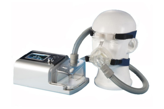
If CPAP/BiPAP is unsuccessful or if whumpee progresses to respiratory failure, they will likely have be intubated. In this case, they will be put under heavy sedation with sedatives (like midazolam [Versed]) and paralytics (like vecuronium) so that they will not be distressed by the breathing tube. They will be weaned from the ventilator when they can maintain their own oxygen saturation.
After treatment in the ER, whumpee will either be admitted to the medical-surgical unit (if they are stable) or the ICU (if they are on CPAP/BiPAP or a ventilator, or in they are unstable in any way). Nurses will round every 4 hours on the med-surg unit and every 2 hours on the ICU to take vitals, give meds, and check the whumpee's respiratory status. If whumpee is conscious and not on CPAP/BiPAP, they will be encouraged to drink plenty of fluids, eat high-calorie-high-protein meals, and walk around the unit if they can do so safely. They will also continue to receive narrow-spectrum antibiotics, pain medications based on their pain level (1-3/10 pain: acetaminophen or ibuprofen; 4-7/10 pain: Percocet or codeine; 8-10/10 pain: morphine or oxycodone), and medications to help them cough up mucus (like guaifenesin). They will likely also be encouraged to use an incentive spirometer, which you can find information on in this post.
Assuming they are not experiencing respiratory distress or failure, whumpee with probably be discharged from the med-surg unit after 2-3 days but will continue taking oral antibiotics for 1-2 weeks. If they are on the ICU, they will likely be transferred to the med-surg unit once they are stable for 24 or so hours of observation, after which they will be discharged to continue taking antibiotics at home. They will be scheduled for a follow-up appointment a week after discharge.
Happy whumping!
5 notes
·
View notes
Text
Is kidney transplant necessary only if you have kidney disease?
Kidney disease is a serious health condition that affects thousands of people every year. Although it can be controlled with treatment, in some cases kidney transplant therapy becomes necessary. But how do you know when such an intervention is needed? In this blog, we will explore what kidney transplant therapy involves when it is needed and what are the alternatives to this treatment. If you are looking for the Best Nephrology Unit in Bardhaman, then you should look into it. Then you can easily be aware of the treatment.
What is kidney disease?
Kidney disease refers to a condition that gradually reduces the function of the kidneys. Since the kidneys play an important role in filtering waste, maintaining electrolyte balance, and regulating blood pressure, any impairment of the kidneys can lead to serious health complications.
Some common types of kidney disease include -
Caused by diabetes, high blood pressure, or genetic predisposition, this condition causes a gradual decline in kidney function.
This condition is caused by sudden damage to the kidneys due to infection, medication or serious injury.
CKD this is the final stage. At this stage, kidney function is almost completely lost. This is when dialysis or kidney transplant therapy becomes necessary.
Types of Kidney Transplant Therapy -
Dialysis and Kidney Transplant Therapy There are two primary types of dialysis and kidney transplant therapy. - Dialysis and kidney transplantation.
Hemodialysis –
A machine is used to filter the blood and remove waste and excess fluid. This process is usually done at least three times a week in a hospital or dialysis center.
Peritoneal dialysis -
Uses the lining of the abdomen (peritoneum) to filter waste through a special fluid that is inserted into the abdomen. This procedure can be done at home, which is more convenient.
Kidney Transplant -
A surgical procedure in which a diseased kidney is replaced with a healthy kidney from a donor. Although a transplant provides a long-term solution, not all patients are eligible due to age, overall health, or lack of a compatible donor.
Why choose Best Nephrology Unit in Bardhaman to choose between dialysis and kidney transplant?
Deciding between dialysis or a kidney transplant depends on several factors.
Dialysis is an immediate solution for those who are not suitable for surgery.
Although a kidney transplant provides a permanent solution to dialysis, the patient will need lifelong medication and regular medical follow-up.
However, if you are unsure about the best option for your condition, consult Diplomat Nursing Home.
If you want to get advanced nephrology care with state-of-the-art diagnostic facilities, then Diplomat Nursing Home is the best place for you as the Best Nephrology Unit in Bardhaman. If your loved one is struggling with kidney disease, then definitely contact us, don't wait for the symptoms to get worse.
0 notes