#Cell wash centrifuge
Explore tagged Tumblr posts
Visit Tumblr Blog
Explore Tumblr blogs with no restrictions, modern design and the best experience.
Last Seen Tumblr Blogs


Fun Fact
The Tumblr app for Google Glass was released on May 16, 2013.
Text
Cell Washing Centrifuge
Labtron Cell Washing Centrifuge is a high-performance serofuge designed for safe, reliable cell-washing procedures. With a max speed of 4000 rpm, capacity for 12 × 7 ml tubes, and a time range of 0 to 99 minutes, it ensures precise results (± 20 rpm speed accuracy). Its LED display offers user-friendly operation and easy monitoring, delivering consistent blood cell washing for up to 12 tubes per cycle.

#centrifuge machine#centrifuge 12 tubes#centrifuge machine price#centrifuge washing machine#laboratory centrifuge#cell wash#centrifuge unit#centrifuge equipment#cell washing centrifuge
0 notes
Text
Cell Washing Centrifuge

A cell washing centrifuge is a specialized centrifuge used in biological and medical laboratories for the purpose of washing cells. Cells need to be washed in various experimental procedures to remove contaminants, residual media, or unwanted substances before further analysis or experimentation.Cell washing centrifuges are crucial tools in various fields such as cell biology, immunology, and molecular biology.Some advanced models may offer programmable settings for precise control over centrifugation parameters.
0 notes
Text
Cell Washing Centrifuge
The Cell Washing Centrifuge is a specialized laboratory instrument designed for the efficient and gentle washing of cells, typically used in biomedical research, clinical diagnostics, and pharmaceutical development. It facilitates the separation of cells from unwanted substances or solutions, such as media, buffers, or contaminants, by utilizing centrifugal force.

0 notes
Text

Max. Speed=4000-rpm; Max. RCF=2000-times-g; Max. Capacity=12-times-7-ml; Time Setting Range=0-99-min; Speed Accuracy=plusmn-20-rpm. Shop Online at Labtron.us
0 notes
Text
0 notes
Text
Simplifying Monocyte Isolation: How PluriBead Enhances Precision in Cell Separation
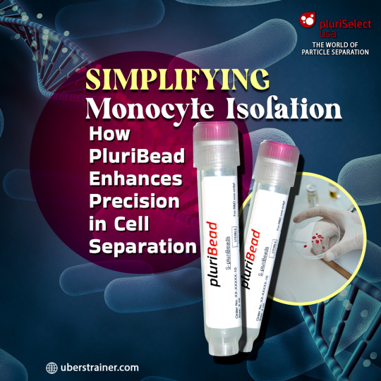
Efficient cell separation is crucial for isolating specific cell types like monocytes, especially in research and clinical applications. Traditional methods often involve multiple steps, expensive equipment, or risk damaging fragile cells. PluriBead revolutionizes this process by offering a magnetic-free, straightforward, and gentle approach to isolating target cells. This article explores how PluriBead simplifies monocyte isolation while enhancing precision in particle separation techniques.
What Is PluriBead Technology?
PluriBead is an innovative cell separation tool designed to isolate target cells without the need for complex equipment or extensive sample preparation. It uses antibody-coated beads to capture specific cells, which are then separated through a simple sieving process using a strainer cascade. The unwanted cells pass through the sieve, while the desired cells remain attached to the pluriBeads for further detachment and collection.
Key Advantages:
No Sample Preparation: PluriBead eliminates the need for gradient centrifugation or erythrolysis.
Versatility: It can process various sample materials, including whole blood, buffy coat, and tissue homogenates.
Rapid Results: Isolation can begin in as little as 5 minutes.
Gentle Process: Ensures high viability and yield, reducing the risk of damaging cells.
How Does PluriBead Work?
Step 1: Mixing
PluriBeads, coated with target-specific antibodies, are added to the sample material. This mixture is gently incubated, allowing the target cells to bind to the beads.
Step 2: Separation Using a Strainer Cascade
The mixture is passed through a pluriStrainer cell sieve. While the unwanted cells run through, the bead-bound target cells remain on top. This process is efficient and leverages particle filtration to achieve high precision.
Step 3: Detachment
Target cells are detached from the beads using a detachment buffer. The detached cells are then washed into a fresh collection tube, leaving the beads behind.
Specialized Features of PluriBead
Cell Enrichment: PluriBead isolates cells with high purity, ensuring that only the desired cell types are collected.
Parallel Cell Isolation: With the pluriBead cascade, researchers can separate two distinct cell types from a single sample.
Sequential Isolation: Up to six different cell targets can be separated sequentially from one sample, making it a versatile solution for complex research needs.
Multiple Applications: Beyond cell isolation, PluriBead can be used for viruses, bacteria, proteins, and RNA/DNA enrichment.
Choosing the Right PluriBead Size
PluriBead is available in two bead sizes:
S-PluriBead: Ideal for isolating small numbers of target cells from large volumes, such as circulating tumor cells (CTCs).
M-PluriBead: Suited for larger target cell populations in smaller sample volumes, such as buffy coat preparations.
Why PluriBead Excels in Particle Separation Techniques
PluriBead’s design eliminates the need for centrifugation and magnetic equipment, making it highly accessible for labs with limited resources. Its reliance on a simple particle filtration mechanism ensures accuracy and reproducibility while maintaining the integrity of fragile cells.
Conclusion
PluriBead’s straightforward workflow, versatility, and ability to achieve high-purity results make it a game-changer in cell separation. Whether isolating monocytes from whole blood or enriching specific cell types for advanced studies, PluriBead delivers precision and efficiency.
In conclusion, cell separation using PluriBead is a cost-effective, reliable, and user-friendly method for researchers and businesses seeking high-quality cell enrichment. Its flexibility across applications and species makes it a valuable tool for advancing cell-based research and therapeutic development.
0 notes
Text
Cell Washer Centrifuge LT-CWC1010

Labtro Cell Washer Centrifuge LT- is an advanced system for serological testing, operating at up to 4000 rpm. Featuring an LED display for speed and time control, it uses SERO and HLA rotors for lymphocyte and erythrocyte washing. Its flexible axle-driven system ensures precise and efficient performance.
0 notes
Text
0 notes
Text
Cell Washing Centrifuge LMCW-A100
Labmate Cell Washing Centrifuge efficiently extracts and cleans blood components at speeds up to 3000 rpm, with a 12-tube capacity and max RCF of 1000×g. Featuring an angle rotor, it’s ideal for both tubes and plates, offering 10 adjustable speed levels for precision control.

0 notes
Text
The Biological Basis of PRP Hair Treatment: An Insight into Hair Restoration

Hair loss is a common challenge that affects men and women of all ages, often leading to decreased self-esteem and frustration. Medical advancements have brought about various treatments to address hair thinning and balding. Among these, PRP (Platelet-Rich Plasma) therapy stands out as a popular technique for promoting hair regrowth due to its effectiveness.
For anyone considering PRP in Abu Dhabi, understanding the science behind this treatment can be beneficial. This blog explores the biological foundation of PRP hair therapy, its benefits, the treatment process, and why it has gained popularity among those seeking hair restoration in the region.
Understanding Platelet-Rich Plasma (PRP)
PRP therapy is a branch of regenerative medicine that utilizes the body's natural healing abilities to support tissue repair and growth. PRP is derived from a patient’s own blood, processed to isolate concentrated platelets. These platelets contain growth factors that play essential roles in cell regeneration, blood vessel formation, and tissue remodeling.
For hair restoration, PRP targets hair follicles, stimulating them to re-enter the active growth phase. Injecting PRP into areas with thinning hair can help improve hair density and reduce hair loss by invigorating dormant hair follicles.
The Science Behind PRP Therapy for Hair Loss
Hair growth occurs in cycles, consisting of three main phases: the growth (anagen) phase, the transitional (catagen) phase, and the resting (telogen) phase. Hair loss can happen when follicles enter the telogen phase prematurely or skip phases in the growth cycle due to factors like hormones, genetics, or aging.
PRP therapy introduces a concentrated supply of growth factors directly to the scalp, encouraging hair follicles to re-enter the growth (anagen) phase faster. Key growth factors in PRP, including platelet-derived growth factor (PDGF), transforming growth factor (TGF), and vascular endothelial growth factor (VEGF), play an important role in stimulating hair follicle activity.
How PRP Promotes Hair Growth:
Cell Proliferation: PRP improves the vitality of dermal papilla cells, which are essential for hair follicle development and growth. This cell activity helps generate thicker, healthier hair strands.
Angiogenesis: Growth factors in PRP also boost hair density by encouraging new blood vessel formation, improving circulation and nutrient supply to hair follicles. This extended supply of nutrients keeps hair in the anagen phase longer, reducing shedding.
Collagen Production: PRP therapy enhances collagen production in the scalp, providing structure and strength to hair follicles. Increased collagen helps make hair thicker and less prone to breakage.
DHT Inhibition: PRP may also counter the effects of DHT, a hormone linked to hair follicle shrinkage and pattern baldness, potentially slowing hair loss and extending growth cycles.
The PRP Procedure for Hair Restoration
PRP therapy for hair loss is a minimally invasive, non-surgical procedure, typically involving the following steps:
Blood Draw: A small sample of the patient’s blood is drawn.
Centrifugation: The blood is then spun at high speed in a centrifuge to separate its components, isolating the PRP from other blood fractions.
PRP Preparation: The plasma with the highest platelet concentration is collected and prepared for injection.
Scalp Injections: PRP is then carefully injected into targeted scalp areas where hair thinning or loss is most evident. While not highly invasive, some patients may experience mild discomfort during injections.
Post-Treatment Care: Patients may be advised to avoid washing their hair for a day or two or follow other specific aftercare instructions as provided by their doctor.
To achieve the best results, PRP therapy often requires multiple sessions. Most patients undergo three to four treatments spaced several weeks apart, with follow-up sessions every six to twelve months to maintain results.
Who Can Benefit from PRP Hair Treatment?
PRP therapy is suitable for individuals experiencing hair loss, particularly those with early-stage hair thinning or conditions like androgenetic alopecia, commonly known as male and female pattern baldness. PRP is an excellent option for those who prefer a non-surgical approach to hair restoration and seek natural treatment methods.
It is important to note that PRP is not a one-time solution for hair loss; ongoing sessions are necessary to maintain results. PRP may not be recommended for individuals with blood disorders or autoimmune diseases.
Read more
0 notes
Text
CQ10 Coenzyme Production Bacteria Screening
Coenzyme Q10, chemical formula C₉HoO₄, also known as ubidecarenone and ubiquinone, is a fat-soluble quinone compound containing a side chain consisting of multiple isoprenoid units connected to the parent nucleus of p-benzoquinone. The molecule contains a side chain of multiple isoprene units connected to the p-benzoquinone nucleus, which has 10 isoprene units. Ubiquinone is involved in energy production and activation in human cells.

Coenzyme Q10 is a natural antioxidant and activator of cellular metabolism. It prevents the oxidation of LDL and cholesterol and reduces the incidence of coronary atherosclerosis. It is used clinically for heart disease, diabetes, cancer, acute and chronic hepatitis, Parkinson's disease, and other diseases, to improve immunity and treat immune system disorders.2 Recent studies have found that Coenzyme Q10 has anti-aging effects. Coenzyme Q10 is produced by plant and animal tissue extraction, chemical synthesis, plant cell culture, and microbial fermentation3 .

Coenzyme Q10 is characterized by the Craven's color test, the Wiss-Brubacher method and thin-layer chromatography. For quantitative analysis, visible spectrophotometry, mass spectrometry (MS), infrared (IR) and ultraviolet (UV) absorption spectrometry (UVAS), optical density analysis (ODA), spectrofluorimetry (SF), and high-performance liquid chromatography (HPLC)l- were used. In this experiment, the content of coenzyme Q10 in Saccharomyces cerevisiae was determined by visible spectrophotometry and inverse high-pressure liquid chromatography (HPLC), and the coenzyme Q10-producing strains of Saccharomyces cerevisiae were screened.

1 Materials and Methods
1.1 Test Materials
1.1.1 Test Strains
Yeast strains from the library were selected as the starting strains for coenzyme Q10 production, as shown in Table 1.
1.1.2 Test medium. Wort 100mL, glucose 10 g, peptone 5g, yeast paste 3g, K₂HPO₄0.5 g, KH₂PO₄0.5 g, MgSO₄-7H₂O 0.3g, pH value 6.0, fixed 1000 mL.
1.1.3 Instruments and Reagents
721 spectrophotometer, ethyl cyanoacetate (analytically pure), anhydrous ethanol (analytically pure), 0.5% potassium hydroxide ethanol solution (weigh 1g of potassium hydroxide (analytically pure) dissolved in 200 mL of ethanol (95%)), coenzyme Q10 standard (Sigma C9538-coenzymeQ10 minium98%HPLC). ).
Table 1 Coenzyme Q10-producing Departure Strains
1.2 Test Methods
1.2.1 Shake Flask Culture, Isolation of Bacteria and CQ10 Coenzyme Extraction
Take 500 mL triangular flask, pour 100 mL of medium, sterilized and inoculated according to 2% of the volume, 200 r/min shaking bed, 30 ℃ shaking culture for 72h, fermentation liquid 3000 r/min centrifugation for 20 min, collect the bacterial body, washed with sterile water twice, centrifugation, to get the yeast bacterial body, weighed. Take 5g of fermented bacteria, transfer to 500 mL round bottom flask, add antioxidant, methanol, sodium hydroxide, distilled water and mix well, reflux at 90 ℃ for 45 min, cool down and add 40 mL of petroleum ether, extract in 2 times, refrigerate at 4 ℃ overnight, remove precipitated proteins and lipids, use rotary evaporator to evaporate out the petroleum ether, and dissolve the yellow oily substance at the bottom of the flask in anhydrous ethanol to be determined.
1.2.2 Visible Colorimetric Standard Curves
Prepare 0.2 mg/mL anhydrous ethanol solution of CQ10 Coenzyme standard, according to the data in Table 2, take different volumes of anhydrous ethanol solution of Coenzyme Q10 standard, add ethyl cyanoacetate and 0.5% potassium hydroxide ethanol solution of 1 mL each, and make a volume of 25 mL, stoppered, shaken, and placed in a dark place at room temperature of 20~30 ℃ for 35 min, and then determine the absorbance at 620 nm (OD) by 721 spectrophotometer, and plot the standard curve with the absorbance and the concentration of Coenzyme Q10. The absorbance at 620 nm was measured by 721 spectrophotometer, and the standard curve was plotted with this absorbance and the concentration of coenzyme Q10.
Table 2 Data related to standard curve of visible colorimetric method
1.2.3 Determination of CQ10 Coenzyme in Strains by Colorimetric Method
Take 3mL of anhydrous ethanol solution and add 1mL each of ethyl cyanoacetate and 0.5% potassium hydroxide ethanol solution respectively, stopper tightly, shake well, and put it in a dark place at 20~30℃ for 35 min, then measure the OD of the samples at 620 nm by using a 721 spectrophotometer, and then calculate the content of CoQ10 in the samples according to the standard curve plotted by the determination.
1.2.4 Identification of Coenzyme Q10 content by HPLC
Chromatographic conditions: the packing material was octadecylsilane-bonded silica gel, the mobile phase was methanol: anhydrous ethanol (1:1), the flow rate was 1 mL/min, the detection wavelength was 275 nm, the temperature was 35 ℃, and the mass concentration of coenzyme Q10 standard (Sigma C9538-coenzymeQ10 minium 98% HPLC) was 0.2 mg/mL, and the injection volume was 10 μL.
1.2.5 Concentration of Samples by HPLC Method
CQ10 Coenzyme samples with concentration differences were measured by visible spectrophotometry and HPLC, respectively, and the correlation between the OD values obtained by visible spectrophotometry and the values determined by HPLC (peak area of the sample/peak area of the standard) were compared to determine the correlation between the data. The correlation between the OD values obtained by VIS and HPLC (peak area of sample/peak area of standard) was determined and the correlation between the data could be estimated.

2 Results and Analysis
2.1 Visible Colorimetric Method for the Determination of CQ10 Coenzyme
2.1.1 Standard curve
The linearity of the standard curve was good, R²=0.997, which can be used to measure the content of coenzyme Q10 by the visible light colorimetric method (Fig. 1); the regression equation of the standard curve of coenzyme Q10: y=3.9268xox is the mass concentration of coenzyme Q10, and y is the OD value.
Figure 1 Coenzyme Q10 standard curve
2.1.2 Determination of CQ10 Coenzyme in Strains by Visible Light Method
The saponification extraction method was used to isolate coenzyme Q10 from the fermenting organisms, and the results of colorimetric detection of coenzyme Q10 in the samples are shown in Table 3, which showed that the highest coenzyme Q10 yields were obtained from C. tropicalis (C. tropi-calis(cast).Berkhout) B021 and Schizosaccheromyces pombe Lindner B147 with reference to the yield of the organisms and the yield of coenzyme Q10 extracted. Berkhout) B021 and Schizosaccharomyces pombe Lindner (B147) had the highest coenzyme Q10 yields.
Table 3 Content of coenzyme Q10 in different strains of bacteria
2.2 Validation of the HPLC Method for the Detection of Coenzyme Q10
The samples with higher content were verified by HPLC assay. Comparison with the standard profiles (Fig. 2) showed that CQ10 Coenzyme was produced by both Saccharomyces cerevisiae B147 (Fig. 3) and Pseudohyphae tropicalis B021 (Fig. 4).
Peak time//min
Fig. 2 Graphical representation of standardized products
Figure 4 HPLC profile of B021 sample
The CQ10 Coenzyme content in the samples was determined by HPLC using standard concentration and peak area conversion, and the results are shown in Table 4. The results are shown in Table 4. Like the visible light colorimetric method, high levels of coenzyme Q10 were found in both Saccharomyces cerevisiae B147 and Pseudohyphae tropicalis B021, which were identified as the starting strains for the metabolic control breeding technique.
Table 4 Determination of coenzyme Q10 by HPLC method
2.3 Linearity Between Visible Spectrophotometry and HPLC method
There was a correlation between the values obtained by 721 VIS and the HPLC values with a linear equation: y=0.0675x+0.0187 and a correlation coefficient R²= 0.986 (Figure 5).

3 Conclusion and Discussion
In this experiment, the visible spectrophotometric method was chosen to measure the content of CQ10 Coenzyme. According to the principle that ethyl cyanoacetate can replace the methoxy group on the coenzyme Q10 molecule to produce blue color under alkaline condition, we established a colorimetric detection method for coenzyme Q10 with 721 spectrophotometer. This method has two advantages: firstly, it can qualitatively detect whether the strain contains coenzyme Q10 through the color reaction, and qualitatively screen the strains; secondly, it can determine the OD value according to the degree of color development by 721 spectrophotometer, and then comparing with the standard curve to calculate the content of coenzyme Q10, which can be used to quantitatively analyze the yield of the strains under certain conditions. This method can be used to screen the starting strains of metabolic control breeding, both qualitative and quantitative, is a simple and fast means of coenzyme Q10 detection.
This test used alkaline alcohol saponification for the extraction of coenzyme Q10. Organic solvents have also been used in the literature. Due to the cellular structure of Saccharomyces cerevisiae, organic solvent extraction requires wall-breaking. However, it was found that the alkanol saponification method was relatively more effective. The saponification time and temperature affect the yield of coenzyme Q10, and there is a certain loss of coenzyme Q10 during the transfer of the extract, protein and fat removal, and evaporation of the extract. Therefore, the actual coenzyme Q10 content was higher than the measured value. The slightly higher values of coenzyme Q10 determined by the visible colorimetric method for strain screening may be due to the presence of some impurities in the yeast cell extracts, which may have interfered with the results of the assay.
In this experiment, two strains, C. tropical- alis (cast) Berkhout B021 and Schizosaccharomyces pombe Lindner B147, were screened for the production of coenzyme Q10. According to the literature7 , coenzyme Q10 production may be further increased if the cultivation conditions are improved with the addition of suitable precursors for coenzyme Q10 biosynthesis. In addition, it is expected that the yield of coenzyme Q10 can be increased by optimizing the nutrient conditions of the fermentation culture of the strains and using metabolic control breeding techniques, such as I3-9: nutrient-deficient mutation, anti-feedback-regulated mutant strains, selecting nutrient-deficient revertant or conditional mutant strains, to deregulate the key enzymes of the end-products, or constructing engineered strains to select high-yielding strains.
References:
[1] Zhang H, Wu YH. Research progress of coenzyme Q10, a vitamin-like substance[J]. Overseas Medicine Health Branch, 2002,29(6):370-373.
[2] Yang HM. Biological effects and synthesis of coenzyme Q_(10)[J]. Jiangxi Chemical Industry, 2010(3):1-5.
[3] TAO Zhijie, WANG Changling, LI Yan. New progress in the preparation and application of coenzyme Q10[J]. Livestock and Feed Science, 2010(3):9-11.
[4] QIN Shengli, YU Jiansheng. Research on the identification method of coenzyme Q_(10)[J]. Chemical Engineering and Equipment, 2011(7):174-175.
[5] WANG Gen-Hua, QIAN He, XIAO Gang. Extraction and determination of coenzyme Q10 in fermented bacteriophage[J]. Journal of Wuxi Light Industry University, 2003,22(2):59-62.
[6] Chen Yu. Content analysis and process optimization of coenzyme Q10 produced by fermentation[J]. Science and Technology Bulletin: 2002,18(4):333-336.
[7] Liu L, Li G. Selection of optimal conditions for coenzyme Q10 production by fermentation with Cryptococcus yellows yeast [J]. Journal of Dalian Fisheries Academy, 2004,19(3):199-204.
[8] WANG Pu, ZHANG Xiaojun, SHEN Jiajia, et al. Biosynthesis pathway of coenzyme Q10 and optimization of fermentation process[J]. Chinese Journal of Biochemical Pharmacology 2006,27(3):178-181.
[9] WU Zufang, WENG Peifang, LI Yin, et al. Breeding ideas and optimization strategies of fermentation conditions for coenzyme Q10 production[J]. Food and Fermentation Industry, 2001,27(7):49-53.
#cq10 coenzyme #cozyme 10 #cq10
0 notes
Text
PRP Hair Treatment in Delhi: Your Path to Fuller, Healthier Hair
In a bustling city like Delhi, hair loss is a common issue faced by both men and women. Pollution, stress, and a fast-paced lifestyle can take a toll on the health of your hair. If you’re struggling with thinning hair or hair loss, you may have heard about Platelet-Rich Plasma (PRP) therapy, a revolutionary treatment that is gaining popularity for its effective, natural results. In this blog, we’ll explore how PRP hair treatment in Delhi can be your path to fuller, healthier hair.
What is PRP Hair Treatment?
PRP hair treatment is a non-surgical procedure that uses your body’s own platelets to stimulate hair regrowth. Platelets are a type of blood cell that promotes healing and tissue regeneration. In this treatment, a small sample of your blood is drawn, and the platelets are separated from the rest of the blood using a centrifuge. These platelets, now concentrated into platelet-rich plasma, are then injected into your scalp where hair loss or thinning occurs.
How PRP Hair Treatment Works for Hair Regrowth
The platelets in the PRP solution contain growth factors that help repair damaged hair follicles and promote the growth of new hair. When injected into the scalp, these growth factors activate dormant hair follicles, improving hair density, texture, and overall hair health. The treatment enhances the natural hair cycle by prolonging the growth phase, resulting in thicker, stronger hair.
Benefits of PRP Hair Treatment in Delhi
Natural and Safe: Since PRP uses your own blood, the risk of allergic reactions or side effects is minimal, making it a safe treatment for hair regrowth.
Non-Surgical and Minimally Invasive: PRP hair treatment does not involve any incisions or stitches, making it a quick and relatively painless procedure.
Effective for Early-Stage Hair Loss: If caught early, hair loss can be effectively treated with PRP, preventing further thinning and restoring lost hair.
Short Recovery Time: The procedure has minimal downtime, allowing you to resume your daily activities soon after the treatment.
Long-Lasting Results: With regular sessions, PRP therapy can deliver long-lasting improvements in hair thickness and quality.
Who is a Good Candidate for PRP Hair Treatment?
PRP hair treatment is suitable for individuals experiencing thinning hair, receding hairlines, or hair loss caused by conditions like androgenic alopecia. If you are in the early stages of hair loss or want to improve the thickness and quality of your hair, PRP could be a great option. A consultation with a qualified professional in Delhi can help determine if PRP hair treatment is right for you.
The PRP Hair Treatment Process
Consultation: The first step is to consult with a specialist in Delhi who will assess your hair loss and recommend a treatment plan.
Blood Collection: A small amount of blood is drawn from your arm.
PRP Preparation: The blood is processed in a centrifuge to separate the platelet-rich plasma from the rest of the blood.
Scalp Injections: The PRP is injected into targeted areas of your scalp to stimulate hair regrowth.
Post-Treatment Care: You can typically resume normal activities after the procedure, but your doctor may advise you to avoid washing your hair or engaging in strenuous physical activity for a day or two.
PRP Hair Treatment Results
Most patients begin to notice an improvement in hair texture and thickness within a few months of starting treatment. The growth of new hair takes time, but with consistent sessions, PRP therapy can deliver visible, long-lasting results. For optimal results, multiple treatments may be recommended, typically spaced 4-6 weeks apart.
Why Choose PRP Hair Treatment in Delhi?
Delhi is home to some of the best cosmetic and dermatology clinics offering PRP hair treatment. With advanced facilities and experienced specialists, the city provides top-tier care to help you achieve fuller, healthier hair. Choosing to undergo PRP hair treatment in Delhi ensures access to cutting-edge technology and personalized treatment plans, giving you the best chance for success.
Conclusion
If you’re looking for a safe, non-surgical solution to hair loss, PRP hair treatment in Delhi could be the answer you’ve been searching for. With its ability to naturally stimulate hair growth and improve hair quality, this treatment can help you regain your confidence and enjoy fuller, healthier hair.
Are you ready to take the next step toward hair regrowth? Consult a specialist in Delhi today and discover how PRP hair treatment can transform your hair and boost your confidence!
0 notes
Text
Low-Speed Centrifuge 4430×g
Labnics low-speed tabletop centrifuge provides precise separation with a max speed of 4000 rpm, 2250×g RCF and a 6×50 ml capacity. It offers ±10 rpm speed accuracy, memory for programs, stainless steel design and an LCD showing RCF, ideal for cell washing and chemical precipitation.

0 notes
Text
Comprehensive Guide to Cell Isolation Techniques: Methods, Applications, and Advances
Cell isolation techniques are fundamental tools in biomedical research, diagnostics, and therapeutic applications. These methods allow scientists to extract and study specific cell types from a heterogeneous population, enabling a deeper understanding of cellular functions, disease mechanisms, and the development of targeted therapies. This article provides an overview of the most commonly used cell isolation techniques, their applications, and recent advancements in the field.

Download PDF Brochure
1. Density Gradient Centrifugation
Overview:
Density gradient centrifugation is a widely used technique for separating cells based on their size and density. Cells are suspended in a medium and subjected to centrifugation, causing them to migrate to different layers based on their density.
Applications:
Isolation of lymphocytes from blood samples.
Separation of cancer cells from a mixed cell population.
Advances:
Modern advancements include the development of more precise density gradients and the use of automated centrifugation systems, improving the efficiency and reproducibility of cell isolation.
2. Magnetic-Activated Cell Sorting (MACS)
Overview:
MACS utilizes magnetic beads coated with antibodies that specifically bind to target cells. When placed in a magnetic field, labeled cells are retained while non-labeled cells are washed away.
Applications:
Isolation of immune cells, such as T cells, for research and therapeutic purposes.
Separation of stem cells for regenerative medicine.
Advances:
Recent innovations in MACS include microchip-based platforms that allow for high-throughput and automated cell sorting, reducing time and labor.
3. Fluorescence-Activated Cell Sorting (FACS)
Overview:
FACS is a powerful technique that sorts cells based on their fluorescence characteristics. Cells are tagged with fluorescent markers and passed through a laser beam; the emitted light is detected and used to sort cells into different populations.
Applications:
Isolation of specific cell types for gene expression studies.
Sorting of transfected cells for gene therapy research.
Advances:
Recent developments in FACS include multi-parametric sorting, allowing for the simultaneous isolation of cells based on multiple characteristics, increasing the precision of cell separation.
Request Sample Pages
4. Immunomagnetic and Immunodensity Cell Isolation
Overview:
These techniques combine magnetic and density-based methods with antibody labeling to isolate specific cell populations. Immunomagnetic separation uses magnetic beads, while immunodensity separation employs density gradients combined with antibodies.
Applications:
Isolation of rare cell populations, such as circulating tumor cells (CTCs).
Enrichment of specific cell types for downstream analysis.
Advances:
Enhanced antibody specificity and the integration of automation have improved the accuracy and efficiency of these methods, making them more suitable for clinical applications.
5. Microfluidic-Based Cell Isolation
Overview:
Microfluidic technology enables the manipulation of cells in tiny channels, allowing for precise control over cell sorting based on size, shape, and other physical properties.
Applications:
Isolation of single cells for genomic and proteomic analysis.
Sorting of cells in personalized medicine applications.
Advances:
The integration of microfluidics with other technologies, such as real-time imaging and AI-driven analysis, has significantly enhanced the capabilities of cell isolation, making it faster and more efficient.
Conclusion
Cell isolation techniques are crucial for advancing our understanding of biology and improving therapeutic outcomes. As technology continues to evolve, these methods are becoming more precise, efficient, and accessible, enabling researchers and clinicians to explore new frontiers in cell-based research and therapy. Whether it's isolating immune cells for cancer treatment or separating stem cells for regenerative medicine, these techniques are paving the way for groundbreaking discoveries and innovations.
Content Source:
0 notes
Text
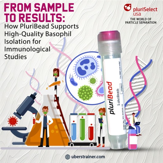
From Sample to Results: How PluriBead Supports High-Quality Basophil Isolation for Immunological Studies
PluriBead provides a streamlined, non-magnetic solution for efficient basophil isolation, simplifying the process and enhancing sample quality through advanced antibody cell separation techniques.
When it comes to high-quality cell isolation, the right technology can make all the difference. For researchers focused on basophil isolation and related immunological studies, PluriBead offers a cutting-edge solution that simplifies and enhances the process. This blog will explore how PluriBead supports efficient basophil isolation using advanced antibody cell separation techniques, delivering reliable results for your research needs.
What Makes PluriBead Stand Out?
PluriBead is a unique cell separation technology that distinguishes itself by working without magnetic components. This non-magnetic approach is designed to streamline the process, making it simpler and more efficient. Unlike traditional methods that require complex procedures or additional equipment, PluriBead offers a straightforward solution: sieve the PluriBeads with bound target cells through a lab cell strainer. The beads with your target cells remain on top, while unwanted cells pass through. After a simple detachment step, your target cells are ready for further analysis.
Key Features of PluriBead for Basophil Isolation
No Sample Preparation Required: One of the main advantages of PluriBead is its simplicity. You can use samples ranging from 200 µl to 45 ml without the need for gradient centrifugation, erythrolysis, or other pre-treatments. This saves time and reduces the complexity of your workflow.
Versatile Sample Materials: PluriBead can handle a wide range of sample types, including whole blood, buffy coat, PBMCs, and various tissues like brain homogenates, spleen, and liver. This versatility ensures that you can adapt the technology to your specific research needs.
High Range of Species: Whether you're working with human, mouse, rat, bovine, canine, or sheep samples, PluriBead accommodates various species. This broad application range is beneficial for comparative studies and cross-species research.
Fast and Gentle Isolation: PluriBead supports fast isolation starting from just 5 minutes. Its gentle approach ensures a high yield of viable cells, minimizing sample requirements and preserving the integrity of your basophils.
Universal Beads for Any Antibody: The PluriBead technology is compatible with any external antibody, providing flexibility in selecting the most suitable markers for your research.
Simultaneous and Sequential Cell Isolation: With PluriBead Cascade straining, you can separate two different cell types simultaneously from a single sample. Additionally, sequential isolation allows you to target up to six different cells from one sample, enhancing your research efficiency.
How PluriBead Enhances Basophil Isolation
Basophils play a critical role in immune responses and are often challenging to isolate due to their low abundance and delicate nature. PluriBead’s non-magnetic cell separation technology is particularly advantageous for isolating these cells. By leveraging PluriBead, researchers can achieve high-purity basophil samples without the complications associated with magnetic bead-based methods.
1. Simple and Effective Workflow: The PluriBead process is streamlined. Start by labeling your sample with the appropriate antibody. Next, incubate the sample to allow binding of the target basophils to the beads. During isolation, the beads with the bound basophils are retained, while the non-target cells are removed. Washing and detaching the basophils from the beads completes the process, providing a ready-to-use sample for downstream applications.
2. Enhanced Sensitivity and Specificity: By avoiding the need for complex pre-treatments and using a non-magnetic approach, PluriBead minimizes potential sources of error and maximizes sensitivity. This ensures that you obtain high-quality basophil isolates, which is crucial for accurate immunological studies.
3. Flexible Applications: PluriBead’s ability to handle various sample types and its compatibility with different antibodies make it a versatile tool for a wide range of applications. Whether you are studying basophils in blood samples or tissue homogenates, PluriBead adapts to your needs.
Conclusion
For researchers and labs focused on basophil isolation and immunological studies, PluriBead offers a robust and efficient solution. Its non-magnetic cell separation technology simplifies the workflow, providing high-quality basophil samples without the need for extensive sample preparation. By incorporating PluriBead into your research, you can leverage advanced antibody cell separation techniques and cell separation technology to enhance your studies and achieve reliable results.
Explore how PluriBead can transform your cell isolation processes and support your research endeavors today.
#antibody cell separation#cell separation technology#particle separation techniques#particle filtration#40 um cell strainer#cell enrichment techniques#medical#uberstrainer
0 notes
Text
Types of Laboratory Plasticware and Their Uses: A Comprehensive Guide

Laboratory Plasticware has become an important part of modern research and experiment. These plasticware is made with high quality plastic which are versatile, durable and chemical resistant. In this comprehensive guide we will tell you about the types of glassware and there uses.
Laboratory plasticware refers to the wide range of laboratory containers used in scientific laboratories for mixing , storing and transporting the chemicals from one place to another. Shifting from glass to plasticware offers numerous benefits in terms of safety, versatility and efficiency.
laboratory Plasticware and their uses
A) Plastic Beakers: used for mixing, stirring and short-term storage of liquids. They come in a variety of sizes and are often labeled for volume measurement.

b) Plastic cylinder: designed for precise measurement of the amount of water. It is available in different capacities and it is necessary to ensure the right solution.

c) Reagent bottles: used to store and distribute chemicals and solutions. They often open for easy pouring and are available in a variety of sizes and screw caps.

d) Wash Bottles: Squeeze the vials to clean the glass or add a small amount of solution to the reactions. It usually has a curved opening for direct flow of water.
E) Pipette Stand: Although not containers, these shelves are important for organizing and storing different types of pipes, keeping them clean and easily accessible.

f) Plastic flask: Available in different types like Erlenmeyer flask, volumetric flask and round bottom flask. It is used for mixing, heating and storing water and chemical.

g) Plastic petri dishes: round dishes used for microbial growth or small experiments. It is often used in microbiology and cell culture applications.
h) Test tubes and racks: used for small tests, sample storage and centrifugation. Integrated shelves help you organize and store multiple test tubes.

Materials Used in Laboratory Plasticware
The choice of plastic material depends on the specific application and chemical compatibility. Common ones:
Polypropylene (PP): resistant to many chemicals and suitable for autoclaving.
Polyethylene (PE): flexible and chemical-free, suitable for general use.
Polycarbonate (PC): light and flexible, suitable for visible tests..
Polytetrafluoroethylene (PTFE): Highly resistant to chemicals and non-stick, used for special applications.
0 notes