#metacercaria
Text
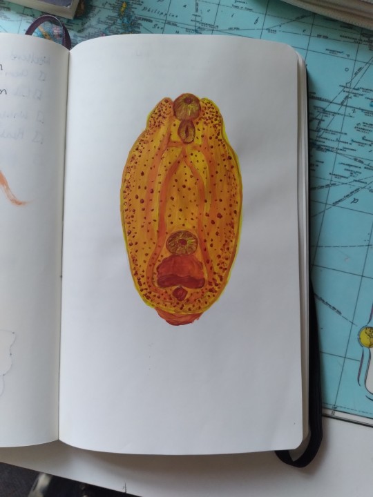
This snail is dead and it's been taken over by a parasite controlling its motor functions & eye stalks, mimicking caterpillars. The parasitic worm wants the snail to be eaten by birds, in whose intestines it will reproduce. 🐌
The snail is infected by a parasitic flatworm called Leucochloridium paradoxum, also known as the green-banded broodsac. This parasite has a complex life cycle that involves two hosts: a land snail and a bird. The parasite’s eggs are released by the bird in its feces, which are then ingested by the snail. The eggs hatch into larvae called miracidia, which develop into a branching structure called a sporocyst inside the snail’s body.
Some of the branches of the sporocyst grow into long tubes that end in a swollen sac, which is called a broodsac. The broodsac occupies one or both of the snail’s eye stalks, where it pulsates and displays bright green and yellow bands. These colors and movements mimic the appearance of a caterpillar, which is a prey item for many birds. The parasite also alters the snail’s behavior, making it more likely to expose itself to sunlight and predators.
The parasite’s goal is to get the snail eaten by a bird, which is its primary host. When the bird consumes the broodsac, it releases hundreds of metacercariae, which are the infective stage of the parasite. The metacercariae travel to the bird’s cloaca, where they mature into adult worms and reproduce sexually. The cycle then repeats itself with the bird’s feces.
The parasite is very harmful to the snail, as it consumes its nutrients, damages its tissues, and reduces its chances of survival. The snail may not be dead yet, but it is essentially a zombie controlled by the parasite. The parasite is also a potential threat to human health, as it can cause skin irritation and allergic reactions if it penetrates human skin. I hope you learnt something new today. 😊🙏
#snail#parasite#worm#leucochloridium#zombie#greenbandedbroodsac#caterpillar#mimicry#schistosomiasis#bilharzia#snailfever
32 notes
·
View notes
Text
Linking metacercariae and adults of Microphallus basodactylophallus (Digenea: Microphallidae), based on larval stages from ctenophores and adult parasites from aquatic birds found in Mexico
http://dlvr.it/T0m2W9
0 notes
Text
Sheep Liver Fluke Life Cycle
1. adult liver fluke lives in sheep liver
2. egg containing a miracidium leaves the sheep’s gut via feces
3. miracidium develops
4. miracidium hatches in fresh water
5. miracidium enters snail
6. becomes a sporocyst with developing rediae
7. rediae develop cercariae
8. cercaria encyst on a water plant as a metacercaria
9. metacercaria is eaten by a sheep
10. adult liver fluke lives in sheep liver
.
Patreon | Ko-fi
#studyblr#notes#biology#biology notes#bio#bio notes#sheep liver fluke#liver fluke#fluke#fluke life cycle#parasites#parasitology#parasite life cycle#miracidium#metacercaria#cercariae#cercaria#vet sci#veterinary science#vet science#veterinary medicine#vet med#vet medicine
6 notes
·
View notes
Text
Yellow Diplostomum metacercariae for when I want to learn about trematodes but also want to do art.
4 notes
·
View notes
Text
OBSERVATIONS ON A NEW XIPHIDIOCERCARIA | UTTAR PRADESH JOURNAL OF ZOOLOGY
Cercaria peterina sp., a Xiphidiocercaria, was discovered in Lucknow on Lymaea auricularia (L.). It grows within the mother sporocyst. The cercaria has been encysted in mosquito and chironomus larvae in the lab. Metacercaria has been described briefly. Experiments with Rana cyanophlyctis and Anatas testudineous on feeding have had disappointing effects.
Please see the link :- http://mbimph.com/index.php/UPJOZ/article/view/947
0 notes
Text
Lupine Publishers | Fasciola Hepatica Antioxidant System: Secretory Excretory System Proteins
Lupine Publishers | LOJ Pharmacology & Clinical Research

Abstract
Fasciola hepatica is a trematode parasite with life cycle complex and as an intermediary it uses different species of snails, the most important is Lymnea truncatula. The definitive hosts being mainly ruminant mammals such as cows, sheep and even humans. The disease they produce is called fasciolosis and is more likely to find in warm to temperate climates with high humidity. It is an important parasitic problem in domestic animals and even in humans. This parasite lives in the bile ducts of several mammals. In order to survive in that environment, the parasite must survive to reactive oxygen species (ROS) of the host and those of its own metabolism. Here we review 3 antioxidant proteins of the parasite secretory excretory system and explain its possible mechanism of action. These proteins are: Thioredoxin peroxidase (TPx), Thioredoxin (TRX) and Thioredoxin glutathione reductase, considered a “general disulfide reductase”. The antioxidant detoxification system of the parasite is thus fully described.
Keywords: Fasciola hepatica; Antioxidant proteins; Secretory excretory system
Introduction
Fasciola hepatica is a hermaphrodite trematode parasite belonging to the platyhelminths’, which in its adult form is flattened, lanceolate, brown and measures approximately 3x1.5cm. It has a conical structure in the front end where it is called suction mouth oral and it has a ventral cup-shaped suction cup surrounded by muscle mass that allows it to adhere to the host (Figure 1). Its life cycle is complex and as an intermediary it uses different species of snails, the most important is Lymnea truncatula, the definitive hosts being mainly ruminant mammals such as cows, sheep and humans (Figure 2). The adult form of the parasite produces 3 to 5 thousand eggs per day which leave in the feces of the definitive host and develop at temperatures of 10 to 30 degrees Celsius in an aqueous environment. They hatch causing larvae called miracidia that look for the intermediate host within which they will go through the phases of sporocysts, redid mother, redid daughter and cercariae. The cercarias leave the intermediaries and encyst in plants as metacercarias waiting to be ingested by who will be their definitive host. The metacercarias are very resistant and can survive up to a year before entering the final host. Inside the definitive host they lodge in the bile ducts. The disease they produce is called fasciolosis and is more likely to find in warm and temperate climates with high humidity. It is an important parasitic problem in domestic animals and humans. They cause inflammation of the bile ducts and fibrosis, hypoalbuminemia, anemia and alteration of certain liver enzymes. Although the parasite lives in an anaerobic environment, oxygen is used for some metabolic processes, such as egg production, which generates many oxidizing molecules. Parasites to prevent damage by these oxidants, both those of their metabolism and those of the host organism’s metabolism, have developed a battery of antioxidant defenses that include enzymes that break down oxides and super oxides [1]. While more levels of defense possess a parasite, greater will be its survival within the host tissues. Helminth parasites in general have at least one of the 3 main antioxidant enzymes that are: superoxide dismutase, catalase and glutathione-dependent enzymes. In Fasciola no catalase has been found and it has low glutathione peroxidase activity [2], so it has other antioxidant enzymes. Fasciola hepatica has the following antioxidant enzymes and proteins, all present in the components of the excretory secretory extract which are part of a parasite detoxification system that allows its survival within the host [3,4]. Thioredoxin peroxidase (TPx), is a protein between 26 and 28 kDa which forms dimers and tetramers due to a cysteine residue conserved in position 47 that forms disulfide bridges and is possibly the primary site responsible for oxidation.
This protein has a protective activity against the inactivation of various enzymes by oxidation systems, but it is different from other TPxs from other parasites because in fasciola it works both in the presence of thiol and ascorbate groups, a relevant functional difference [5]. Another relevant fact and different from the other similar proteins of other parasites is that fasciola TPx has an extracellular location extending its protection against antioxidants to enzymes outside the parasite. The TPx mechanism could intervene in processes of protection of the inactivation of some membrane enzymes or exocellular parasite enzymes, making it possible to survive the host defense mechanisms [6,7]. Thioredoxin (TRX), representative protein of a group of widely distributed proteins that have dithiol-disulfide-oxidoreductase activity, its molecular mass is 12 kDa. Participates in important processes of metabolism and homeostasis. In the presence of TRX, the TPx protein protects enzymes more effectively against oxidation systems that employ DTT and / or ascorbate [6]. It is able to catalyze in vitro renaturation of both denatured insulin and RNase so it could be the natural physiological electron donor of the TPx protein [4]. It participates in the electron transfer pathway through the reversible oxidation of two neighboring cysteines in a biochemical cycle that involves TRX reductase and NADPH [8,9]. Finally, Thioredoxin glutathione reductase, considered as a “general disulfide reductase” is involved in the reduction of exposed disulfide bonds of a variety of proteins and thus the entire system is able to metabolize H2O2 and other alkyl peroxides [10]. With this protein the antioxidant system of detoxification of the parasite is completed whose proposed model is as follows: TPx protects against ROS inactivation in an oxidation system catalyzed by Fe3 + using thiol and / or ascorbate.
Conclusion
The reducing agent is responsible for activating the enzyme by reducing disulfide bridges. Oxidized sulfhydryl groups are regenerated by transferring reducing agents from NADPH to thioredoxin reductase, from it to thioredoxin and finally to TPx as shown in the (Figure 3) [4]. It does not seem logical to think that NAPH acts extracellularly as a source of the necessary reducing power. Therefore, the theory of acting inside the cell is postulated, so that proteins could be secreted in a reduced state. Thus, the parasite defense system against ROS products (reactive oxygen species) generated by the host’s immune response and by the parasite’s own metabolism is described.
https://lupinepublishers.com/pharmacology-clinical-research-journal/pdf/LOJPCR.MS.ID.000123.pdf
https://lupinepublishers.com/pharmacology-clinical-research-journal/fulltext/fasciola-hepatica-antioxidant-system-secretory-excretory-system-proteins.ID.000123.php
For more Lupine Publishers Open Access Journals Please visit our website: https://lupinepublishersgroup.com/
For more Pharmacology & Clinical Research Please Click
Here: https://lupinepublishers.com/pharmacology-clinical-research-journal/
To Know more Open Access Publishers Click on Lupine Publishers
Follow on Linkedin : https://www.linkedin.com/company/lupinepublishers
Follow on Twitter : https://twitter.com/lupine_online
0 notes
Text
Salmon poisoning

Fishing can be wonderful recreation, but sharing the catch with your dog can be an act of kindness that kills.
Salmon Poisoning Disease is a potentially fatal condition seen in dogs that eat certain types of raw fish. Salmon (salmonid fish) and other anadromous fish (fish that swim upstream to breed) can be infected with a parasite called Nanophyetus salmincola. Overall, the parasite is relatively harmless. The danger occurs when the parasite itself is infected with a rickettsial organism called Neorickettsia helminthoeca. It’s this microorganism that causes salmon poisoning.
Dogs and other animals become infected by ingesting trout, salmon, or Pacific giant salamanders that contain the encysted metacercaria stage of the rickettsia-infected fluke. In the dog’s intestine, the metacercarial flukes excyst, embed in the duodenal mucosa, become gravid adults, and transmit the rickettsiae to monocytes-macrophages.
Symptoms
Fever
(Hemorrhagic) Diarrhea
Vomiting
Anorexia
Depression
Swollen lymph nodes (lymphadenopathy)- 60% of cases
Discharge from the nose and eyes
Leukopenia with degenerative left shift (Neutrophilia)
Thrombocytopenia - 94% of cases
Signs appear suddenly, usually 5–7 days after eating infected fish, but may be delayed as long as 33 days, and persist for 7–10 days before culminating in death in up to 90% of untreated animals.
Diagnosis and Treatment

Fluke ova are found on fecal examination in ~92% of cases, which supports the diagnosis. The ova are oval, yellowish brown, rough-surfaced, and ~87–97 × 35–55 μm, with an indistinct operculum and a small, blunt point on the opposite end.
Intracellular organisms have been demonstrated by Romanowsky staining on lymph node aspirates in ~70% of cases.
PCR testing to detect DNA-specific N helminthoeca (or Neorickettsia genus) is recommended for accurate diagnosis.
Serologic testing using the N helminthoeca organism has been developed.
Other causes of fever of unknown origin, generalized lymphadenopathy, vomiting, and diarrhea are differential diagnoses. Currently, the only means of prevention is to restrict the ingestion of uncooked salmon, trout, steelhead, and similar freshwater fish
Sulfonamides are not effective and may exacerbate the clinical disease. Recommended treatment is parenteral oxytetracycline or doxycycline. Oral tetracycline or doxycycline is contraindicated because of impinging GI signs. Animals usually succumb because of dehydration, electrolyte and acid-base imbalances, and anemia. Therefore, general supportive therapy to maintain hydration and acid-base balance, while meeting nutritional requirements and controlling diarrhea, is often essential. Judicious use of whole blood transfusions may be helpful.
1K notes
·
View notes
Photo

RG @svinfectologia RESPUESTA TRIVIA SVI ¿Cuál es el diagnóstico? c) Paragonimiasis. La fibrobroncoscopia mostró área de inflamación de la mucosa, y se encontraron huevos de paragonimus y eosinófilos aumentados en el líquido del lavado broncoalveolar. La serología fue positiva por ELISA. No tenía antecedente de consumo de cangrejos al cocinados o crudos pero le gustaba comer sashimi. Y su condición se controló después del tratamiento con praziquantel. Como una helmintiasis zoonótica, la paragonimiasis se encontró ampliamente en China antes de la década de 1950, pero ahora la enfermedad rara vez se encuentra. Puede ser causada por comer paragonimus metacercarias infecciosas crudas o poco cocinadas (cangrejo y cangrejos de río) o beber agua sin hervir en las áreas de endemicidad. . Los pacientes con paragonimiasis pulmonar típica desarrollan fiebre, dolor en el pecho y síntomas respiratorios, incluida tos crónica con hemoptisis. Sin embargo, las presentaciones clínicas de la paragonimiasis pulmonar son complicadas y con frecuencia son indistinguibles de las de neumonía, tuberculosis pulmonar y cáncer de pulmón. Fuente: J Clin Microbiol. 2014 Feb; 52(2): 710. #respuestatriviasvi https://www.instagram.com/p/B-Gcj4mHN6Q/?igshid=1h4gnmmf2kckt
0 notes
Text
New in Pubmed: Survey of Gymnophalloides seoi Metacercariae in Natural and Cultured Oysters from Several Western Coastal Areas, Korea.
Survey of Gymnophalloides seoi Metacercariae in Natural and Cultured Oysters from Several Western Coastal Areas, Korea.
Korean J Parasitol. 2019 Dec;57(6):705-708
Authors: Chang T, Jung BK, Song H, Cho J, Hong S, Lee KH, Hoang EH, Kang J, Lim J, Lee H, Chai JY
Abstract
Gymnophalloides seoi (Digenea: Gymnophallidae) is a human intestinal trematode contracted by eating raw oysters (Crassostrea gigas) in the Republic of Korea (=Korea). It has been known to be highly endemic in Aphae Island, Shinan-gun, Jeollanam-do (Province). However, recent epidemiological status of G. seoi has not been reported since the 1990s. In this study, we investigated the prevalence of G. seoi metacercariae in natural and cultured oysters collected from 3 islands and 2 coastal areas in western parts of Korea. The oysters were examined using the artificial digestion method followed by stereomicroscopy. The overall positive rate of G. seoi metacercariae in natural oysters was 66.0% (99/150), and the oysters collected from Yubu Island showed the highest infection rate (74.0%). However, the metacercarial density per oyster was relatively low (1.5-2.4 per oyster). By contrast, no metacercaria was found in cultured oysters purchased from 2 coastal areas in Chungcheongnam-do. Thus, we could confirm that natural oysters produced from 3 western coastal islands are infected with G. seoi metacercariae, whereas cultured oysters purchased from 2 coastal areas were free from infection.
PMID: 31914525 [PubMed - in process]
from pubmed: crassostrea gigas https://ift.tt/304YqS1
via IFTTT
0 notes
Link
Biology is really one big horror story. You don't need to look much further than the various types of parasites that drive their hosts into the mouths of hungry predators.
If you're keeping track of these tiny monsters, you should know that there's a species of flatworm that parks itself inside the eyeball of a fish, and controls when its host hides from birds or exposes itself to be eaten - all to benefit its very complicated (and creepy) life cycle.
A team led by the Severtsov Institute of Ecology and Evolution in Moscow, Russia found that a common parasite called an eye fluke, Diplostomum pseudospathaceum, evolved this rather gruesome way of navigating its way through its somewhat complex life cycle.
The fluke relies on three different animals to develop from egg to adult:
-It mates in a bird's digestive tract, where the eggs pass into the water with the bird's faeces
-Larvae hatch from the eggs and seek out a freshwater snail to burrow into, where they mature and reproduce asexually
-The next stage of larvae, free-swimming forms of the parasite called cercariae, leave the snail and then dig their way through a fish's hide for their final journey, which ends in the lens of the animal's eyeball in a stage called metacercariae
-A bird eats the fish, infected eyeballs and all, and the fun begins all over again.
Continue Reading.
#Science#parasites#parasitism#Biology#zoology#eye fluke#fish#ichtyology#stem#sciblr#scienceblr#body horror#flatworm
168 notes
·
View notes
Video
youtube
Learn how to pronounce Metacercaria in English --- METACERCARIA Pronunciation of Metacercaria: /ˌme-tə-(ˌ)sər-ˈker-ē-ə/ noun Definition of Metacercaria: a tailless encysted late larva of a digenetic trematode that is usually the form which is infective for the definitive host ★ http://Learn2Pronounce.com ★ How to pronounce Metacercaria | English pronunciation: https://youtu.be/5K5R0dkz1gM
#how to pronounce How to pronounce Metacercaria | English pronunciation#pronunciation of How to prono
0 notes
Text
clonorchiasis
Introduction
Clonorchiasis, due to Clonorchis sinensis and opisthorchiasis, due to Opisthorchis, occur in Southeast Asia and Eastern Europe
-Clonorchiasis and opisthorchiasis are clinically indistinguishable.
-Humans are infected by eating raw, pickled, frozen, dried, salted, and smoked fish containing the encysted larvae (metacercariae)
-After excystation in the duodenum, immature…
View On WordPress
0 notes
Text
THE INFLUENCE OF METACERCARIAE OF DIPLOSTOMUM (TREMATODA) ON TRE RESPIRATION OF HETEROPNEUSTES FOSSILIS (BLOCH) | UTTAR PRADESH JOURNAL OF ZOOLOGY
In Diplostomum infected H. fossilis, oxygen consumption increases. The rate of growth is determined by the severity of the infection. In the surfacing prevented condition, the percent rise in a particular grade of infection is greater than in the surfacing allowed condition.
Please see the link :- http://mbimph.com/index.php/UPJOZ/article/view/961
0 notes
Text
Parasitoides
"
O parasitismo é um tipo de interação ecológica que ocorre principalmente entre diferentes espécies, onde uma espécie parasita se associa a um ou vários indivíduos hospedeiros, causando-lhes prejuízo. Assim, o parasita pode ser definido como um organismo que obtém recursos através do hospedeiro, provocando danos e reduzindo sua aptidão sem causar, no entanto, morte imediata. Quando esse tipo de interação negativa, ou desarmônica, acarreta diretamente na morte do indivíduo hospedeiro ela passa a ser chamada de parasitoidismo. Os parasitoides são caracterizados por apresentar alta fecundidade, ciclos de vida curto e ainda interagirem de maneira bastante específica com seus hospedeiros.
A maioria dos parasitoides pertencem ao grupo dos insetos, principalmente às ordens Diptera (moscas) e Hymenoptera (vespas), e as fêmeas costumam depositar seus ovos sobre dos hospedeiros. Assim, ao eclodirem dos ovos, as larvas consomem o indivíduo hospedeiro que serve de recurso na fase inicial do desenvolvimento, matando-o muitas vezes antes dele se reproduzir pela primeira vez. Algumas vespas parasitoides pertencentes a subordem Apocrita apresentam um ovipositor utilizado para inserir os ovos no corpo do hospedeiro. Já a vespa da espécie Cotesia congregata deposita seus ovos nas larvas do inseto do tabaco, a mariposa Manduca sexta. Junto com os ovos, essa espécie injeta um vírus que derruba o sistema imunológico do hospedeiro, facilitando a invasão e eclosão dos ovos.
//<![CDATA[ googletag.cmd.push(function() { googletag.display('dfp-arroba-meio1'); }); //]]>
É comum que o parasitoidismo modifique o comportamento da espécie hospedeira, facilitando o desenvolvimento e propagação do parasitoide e também estabelecendo uma relação de alta especificidade. Por exemplo, o verme trematódeo Dicrocoelium dendriticum infecta formigas na fase de metacercaria, atingindo seu cérebro e alterando seu comportamento, fazendo com que a formiga fique em cima das folhas ao invés de se proteger contra predadores e se torne mais fácil de ser ingerida por herbívoros. Nesse caso, os herbívoros como bovinos, ovinos, cervídeos e coelhos são o hospedeiro definitivo do parasitoide.
Classificação
Existem diversas formas de se classificar os organismos parasitoides. Em relação ao local de desenvolvimento, os indivíduos podem se desenvolver na superfície externa do hospedeiro, também chamados de ectoparasitoides, ou no interior do corpo do hospedeiro, conhecidos como endoparasitoides. Já em relação a fase de vida do hospedeiro, os parasitoides podem atacar e se desenvolver em ovos, larvas, pupas, ninfas e também no hospedeiro adulto. Em todos esses casos a exploração do hospedeiro pode acontecer de duas formas, classificando os parasitoides em idiobiontes, que inibem qualquer atividade e desenvolvimento do hospedeiro no momento da parasitação, e cenobiontes, que permitem que o hospedeiro se alimente e se desenvolva, morrendo posteriormente por conta da eclosão das larvas.
//<![CDATA[ googletag.cmd.push(function() { googletag.display('dfp-arroba-meio2'); }); //]]>
Ao se desenvolver em hospedeiros não parasitados, o parasitoide também pode receber o nome de parasitoide primário. Em alguns casos, os parasitoides chamados secundários ou hiperparasitoides parasitam outros parasitoides, e costumam ser ingeridos por organismos já parasitados anteriormente. Por exemplo, as vespas fêmeas da família Trigonalidae ovipositam seus ovos em folhas que servirão de recurso alimentar para lagartas. Ao atingirem o sistema digestivo, perfuram a parede intestinal e procuram por outras larvas parasitoides para se alimentarem.
Controle biológico de pragas
Uma vez que a relação entre parasitoides e seus hospedeiros é bastante específica, é comum que pragas agrícolas sejam controladas naturalmente através do parasitoidismo. Vespas da família Ichneumonoidea e Braconidae parasitam respectivamente lagartas de borboletas e traças, e até mesmo afídeos. Em agrossistemas, como cultivos de café, algodão, soja, sorgo, feijão e trigo, são encontrados dezenas de famílias de parasitoides responsáveis pelo cultivo de pragas. A superfamília mais encontrada costuma ser a Chalcidoidea, uma vez que apresenta elevada diversidade de hospedeiros, como afídeos, moscas, lagartas e cochonilhas.
Referências Bibliográficas:
[1] Begon, M.; Townsend, C. R. & Harper, J. L. Ecology: from individuals to ecosystems. 4 ed. Reino Unido: Editora Blackwell Publishing Ltd, 759p., 2006.
[2] Parasitoid. Disponível em: https://en.wikipedia.org/wiki/Parasitoid.
[3] Souza, L.; Braga, S.M.P; Campos, M.J.O. HIMENÓPTEROS PARASITÓIDES (INSECTA, HYMENOPTERA) EM ÁREA AGRÍCOLA DE RIO CLARO, SP, BRASIL. Arq. Inst. Biol., São Paulo, v. 73, n. 4, p. 465-469, 2006.
The post Parasitoides appeared first on InfoEscola.
InfoEscola https://ift.tt/2VMqSoB
Publicado primeiro em https://www.infoescola.com"
Este conteúdo apareceu primeiro em: https://ift.tt/2w0BWWY
0 notes
Text
Exploring the genetic diversity of Tylodelphys (Diesing, 1850) metacercariae in the cranial and body cavities of Mexican freshwater fishes using nuclear and mitochondrial DNA sequences, with the description of a new species.
http://dlvr.it/Qtk2db
0 notes
Photo

Metagonimus yokagawai (si monster bagi pecinta sushi) menginfeksi manusia👨🏻👧🏻 yang mengkonsumsi ikan 🐟 (terutama ikan mentah) yang mengandung metacercariae. Spesies ini tersebar di Korea🇰🇷, China🇨🇳, Taiwan🇹🇼, Jepang🇯🇵, Rusia🇷🇺, Indonesia🇲🇨, dan Spanyol🇪🇸. Regrann dari @instaparasite . Untuk pembelian buku-buku Analis Kesehatan/ ATLM hubungi 085697529868 (SMS/WA). . #analis_kesehatan #atlm #tlm #medical #laboratory #hematology #analiskesehatan #laboratorium #medis #kesehatan #analis #teknologilaboratoriummedis #teknologilaboratoriummedik #microbiology #lab #parasitology #medicallaboratory #bacteriology (di Toko Buku ATLM dan Analis Kesehatan) https://www.instagram.com/p/BoYbeYMHuqZ/?utm_source=ig_tumblr_share&igshid=11v98dxeih0mo
#analis_kesehatan#atlm#tlm#medical#laboratory#hematology#analiskesehatan#laboratorium#medis#kesehatan#analis#teknologilaboratoriummedis#teknologilaboratoriummedik#microbiology#lab#parasitology#medicallaboratory#bacteriology
0 notes
Last Seen Blogs

addictredcan3
Untitled

bgmguru18
Untitled

cv-rajakulit
leather industries

informasipertanian
informasi pertanian

pinkshastagirl
Pink Shasta Girl